Ultrasound-Guided Drainage
Home » Resources » Ultrasound-Guided Procedures » Ultrasound-Guided Drainage
History
Things To Do With A Needle & A Probe: Ultrasound-Guided Drainage
Basically with sedation and avoidance of vital structures, any cavity or compartmentalized fluid filled or viscous structure may be drained and analyzed with cytology and culture. Pericardiocentesis, thoracocentesis, abdominocentesis, and cystocentesis are the traditional ultrasound guided drainage procedures that have been performed most historically.
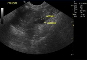
US guided abscess drainage and antibiotic injection into various organs has been a technique utilized frequently on our team with solid levels of success and is currently a subject of study. No evidence of complications has been observed. The reasoning lies in the fact that abscesses tend to have a granulation bed or organization of tissue that walls them off from the tissue circulation. The body’s own immune system attempts to sectorialize the abscessing pathology to minimize systemic affects. This causes reduced blood flow into the abscess or necrosis itself that may be evidenced on power Doppler assessment or the abscess and surrounding tissue.
If there is no blood flow to the lesion then its reasonable to assume that even endovenous medications and even the body’s own immune elements will have difficulty reaching the sectorialized pathology. Therefore, a clinical sonographer armed with a 2 inch 14-16- IV catheter may drain an abscess or infected cyst in any organ and follow the drainage procedure with injection of a body weight dose of antibiotic directly into the lesion with a 22-gauge needle. (prostatic abscess injection.avi)
This has been performed with success and without complication in our experience in the liver, pancreas, omental abscesses, lung, prostate, and spleen. We do not recommend this in the kidney for concern for renal toxicity. Enrofloxacin or other quinolones have been usually the antibiotic of choice in these procedures but cephalosporins and ampicillin have also been utilized. There is no way of knowing at the time of the drainage procedure whether the abscess is septic or not or if the injected antibiotic is actually specific for the potential bacteria involved.
However, considering the spectrum of antibiotics that may be injected endovenously (criteria for selection of the antibiotic), we tend to utilize the antibiotic that likely would have an effective spectrum against the usual pathogens that may dwell in the affected organ.
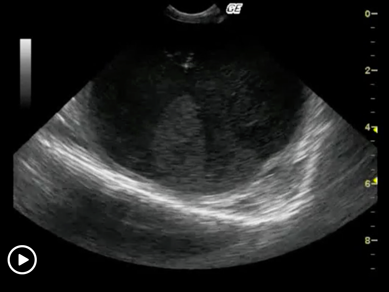
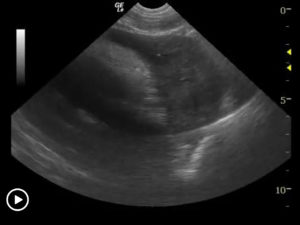
Example of adbsess drainges are demonstrsated in the videos below:
More Ultrasound-Guided Procedures
Bubble Study
The reason to perform a bubble study is to confirm the presence of shunting of blood from venous to arterial flow such as a Reverse VSD or a Reverse PDA.

Gallbladder Motility Study
NPO overnight. Time 0 min: Measure GB in long axis and short axis from subxiphoid approach and right intercostal approach. The proper position is maximum
Intraoperative Ultrasound
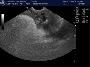
We developed this technique to be used on any abdominal organ but is especially effective in case of infiltrative, focal and multifocal GI lesions.
Lindquist Compression Technique For Thoracic FNA
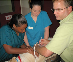
This technique often makes inaccessible thoracic lesions accessible.
Traumatic Catheterization Procedure
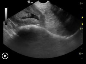
Sedation Use largest open-end rigid polypropylene urinary catheter cut diagonally to create a sharp edge like a needle bevel. Lube heavily and pass catheter retrograde

UGELAB for Transitional Cell Carcinoma in the Dog
Dr. Dean Cerf, Ridgewood Veterinary Hospital NJ and Dr. Eric Lindquist, SonoPath.com For More Information Contact Dr. Dean Cerf at Ridgewood Veterinary Hospital, Ridgewood, NJ,
Ultrasound-Guided Lymph Node Culture
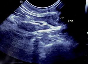
This type of lymph node sampling is helpful in determining infected lymph nodes vs. lymphoma. Supplies: 6 cc Luer lock syringe 1cc of saline 2
Ultrasound-Guided Pericardiocentesis
History Pericardial effusions typically cause some level of cardiomegaly on radiographs but note poor vascular volume in the pulmonary artery and vein. Muffled heart sounds

Ultrasound-Guided Sampling Procedures
Description Ultrasound-guided needle sampling is frequently used in the diagnostic evaluation of patients. Fine needle aspiration has the advantage of requiring minimal or no sedation
