Left Adrenal Mass With Caval Invasion: Our Case Of the Month February 2024
SonoPath’s New Jersey Mobile and SDEP® Certified clinical sonographer Kenneth Leal, DVM, captured the high-end, diagnostic images for this case with detailed interpretation by Eric Lindquist, DMV, DABVP, Cert. IVUSS. Many thanks to Dr. Adam Boe of Smithfield Animal Hospital located in East Stroudsburg, PA for providing excellent medical management of this case and to the compassionate staff for giving the best care for this big fellow.
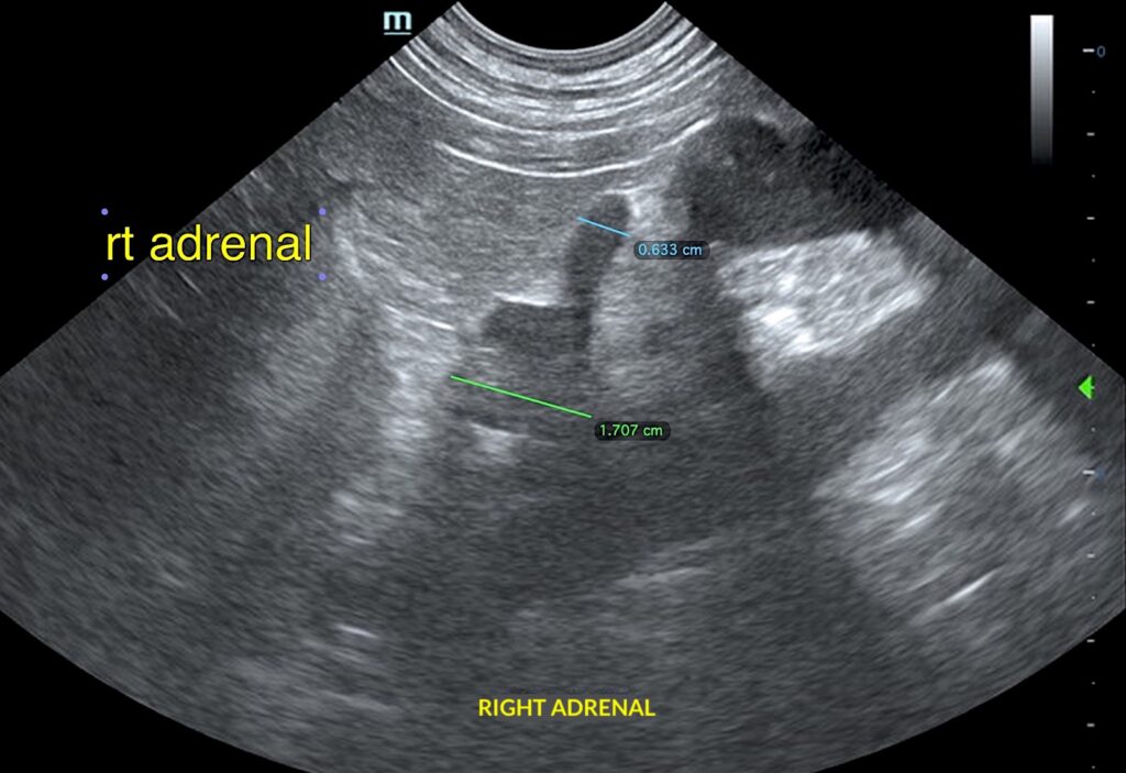
Ultrasound Findings
Echo: Tachycardia and volume contraction of the heart, no evidence of structural disease.
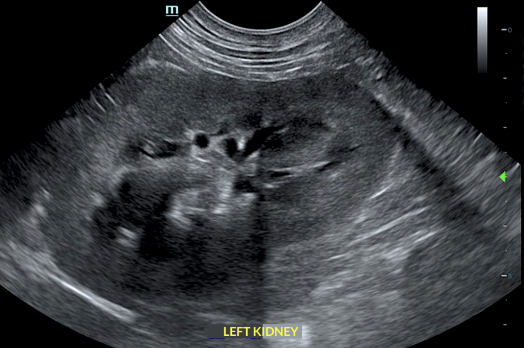
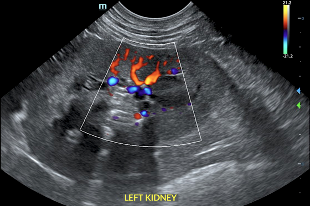
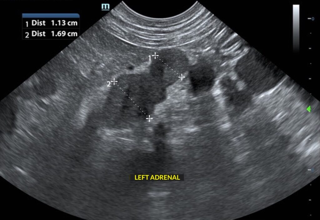
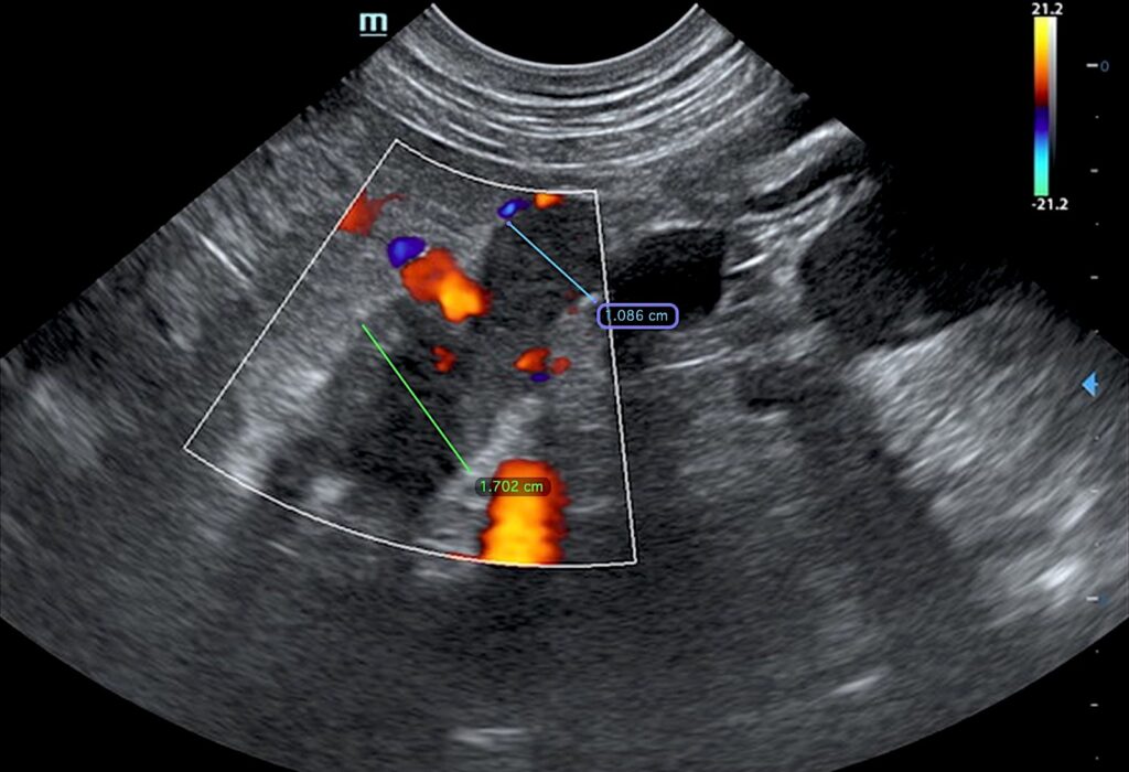
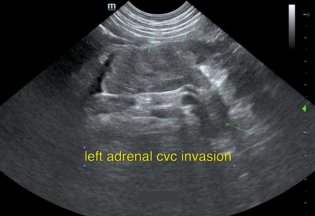
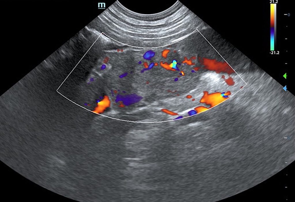
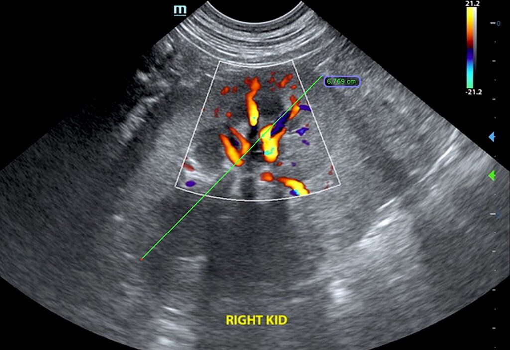
Abdomen: Left adrenal mass invasion – pheochromocytoma suspected, carcinoma less likely. Slightly heterogeneous right adrenal gland. BPH prostate. Age related renal, splenic, and hepatic changes.
Diagnosis
Strong concern for early phrenic vein invasion. Pheochromocytoma, carcinoma, hyperplasia with possible thrombosis are all potentials.
Interpretation of the Findings
No evidence of primary cardiac disease. Surgical consultation is warranted if the surgeon is prepared for left adrenalectomy with 4.0-4.5 cm of caval invasion.
HISTORY: A 10-year-old intact male Pit bull terrier was presented for repeatedly elevated UPC with no other significant symptoms. UA: protein 500, UPC > 5.58. An abdominal and cardiac ultrasound was performed.
Further Recommendations
Referral for surgical consultation was discussed with the owners. At this time, the owners are choosing to manage the patient’s symptoms with medications and will closely monitor him for any new symptoms. Following the ultrasound evaluation, the owners did report that the patient is PU/PD. Concern for Cushing’s disease was conveyed to the owners with a recommendation for an LDDST test to provide more information about the status of how the adrenal glands are functioning. There is no further follow-up at this time, but this case will be updated if additional information becomes available.
Learn More About Adrenal Glands
Continuing Education: Sonographic Adrenal Pathology Gain a better understanding of when adrenal changes are clinically pertinent from a clinical & sonographic perspective. Learn when sonographic changes are simply age-related and have no bearing on case work-up.
From the Merck Manual, Veterinary Manual: “Disorders of the Adrenal Glands in Dogs” By David Bruyette, DVM, DACVIM, Anvive Lifesciences, INC. The adrenal glands are located just in front of the kidneys. The adrenal gland has 2 parts—the cortex and the medulla. The adrenal cortex is subdivided into 3 layers, and each layer produces a different set of steroid hormones. The outer layer produces the mineralocorticoids, which help to control the body’s balance of sodium and potassium salts. The middle layer produces glucocorticoids, which are involved in metabolizing nutrients as well as in reducing inflammation and immune responses. The inner layer produces sex hormones such as estrogen, progesterone, and androgens. The adrenal medulla plays an important role in response to stress or low blood sugar (glucose). It releases epinephrine (sometimes also called adrenaline) and norepinephrine, both of which increase heart output, blood pressure, and blood glucose, and slow digestion. Read the full article By David Bruyette, DVM, DACVIM, by clicking HERE.