
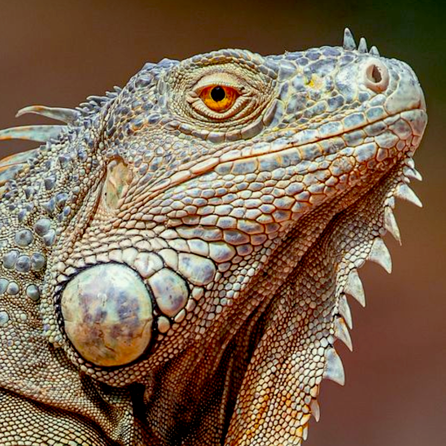
Teamwork makes the dream work! We would like to give credit to Paws, Wings & Scales Animal Hospital who managed this challenging case for their patient, a 17-year-old iguana. Dr. Stancel provided compassionate care and fast treatment for this case. Based on the findings of the ultrasound, Dr. Stancel was able to go immediately into exploratory surgery. Thank you to Dr. Stancel , technician Melissa, and the whole team at Paws, Wings & Scales Animal Hospital, for taking wonderful care of this patient. Shari Reffi, CVT, SDEP® Certified Clinical Sonographer captured the high-end diagnostic images for this case with interpretation and medical recommendations provided by Dr. Eric Lindquist DMV, DABVP, Cert. IVUSS.
ULTRASONOGRAPHIC EXAMINATION OF THE ABDOMEN
The liver revealed a large 5.9 cm cyst. The liver parenchyma was heterogenous. Very full stomach with hard shadowing content that seems excessive for this patient. The visible intestine appeared unremarkable. Uniform curvilinear patterns were maintained.
Slight free fluid was noted around the liver cyst. This is likely owing to potential leakage from the cyst.
SONOGRAPHIC DIAGNOSIS
Liver cyst.
Cranial mediastinal cyst measuring 5.0 cm, with pleural effusion. Potential cyst rupture.
Potential for gastric impaction.
Slight free fluid.
INTERPRETATION OF THE FINDINGS & FURTHER RECOMMENDATIONS
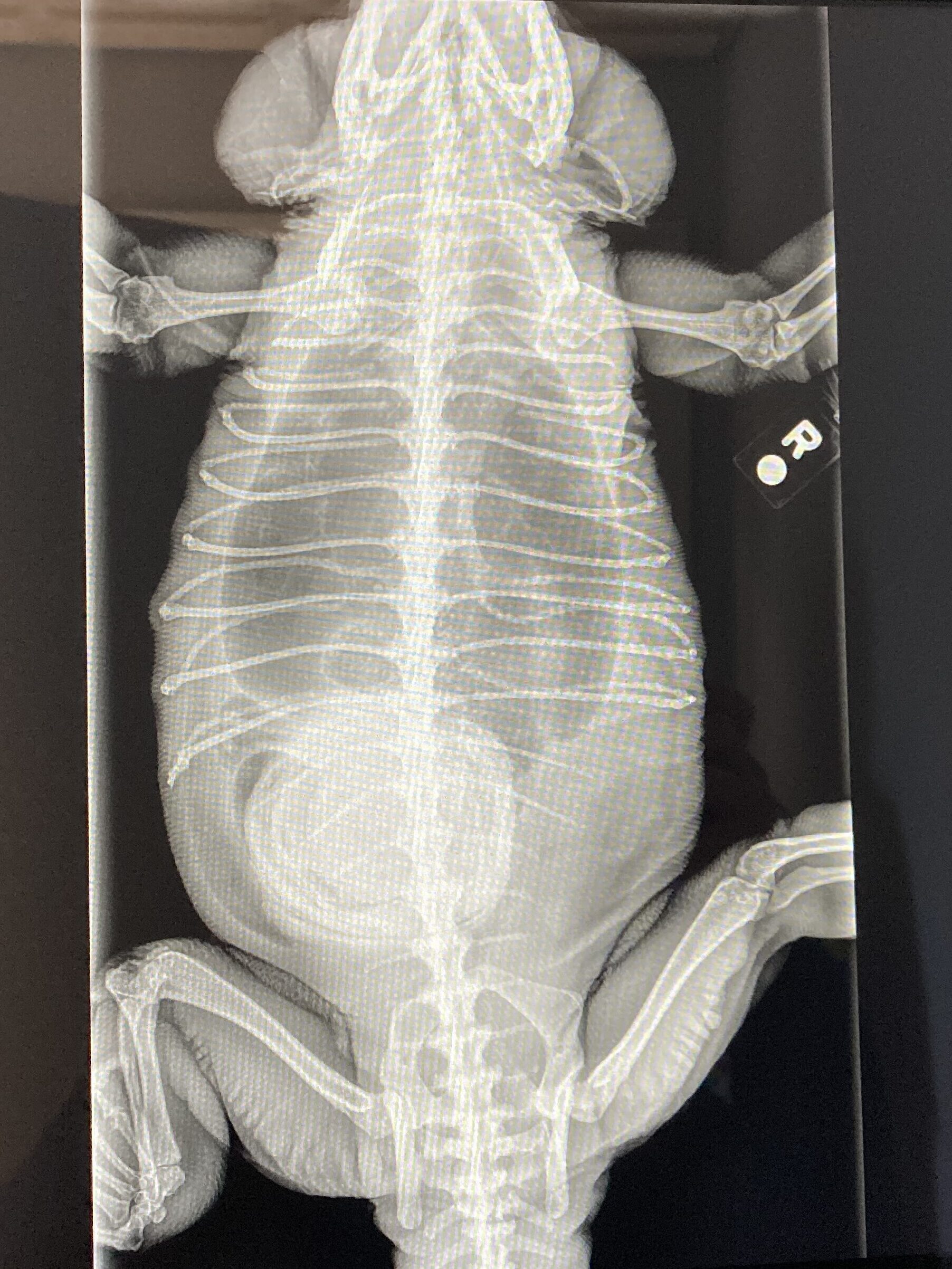
Given the radiodense structure in the cranial abdomen, as well as the pleural effusion, gastric impaction and leakage from the two cystic structures and potential underlying neoplasia are the primary concerns. Exploratory surgery may be necessary to evaluate as well as to perform a cystotomy.

HISTORY:
Owner reports pet was acting odd, fell face-first the night before. PE: Pet pale, palpable mass. No current medications.


CURRENT CASE STATUS:
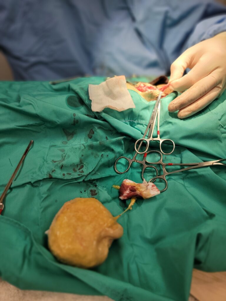
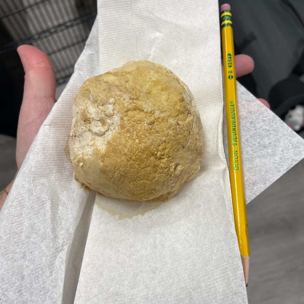
Based on the findings of the ultrasound, Dr. Stancel’s team took the patient in for immediate exploratory surgery and performed a cystotomy. A massive urinary bladder calculus was excised, and at last update the patient is recovering well. No further updates regarding the hepatic cyst or mediastinal cyst were available at the time of publication.

Did you know?
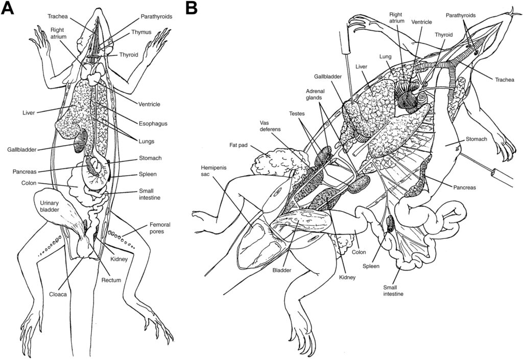
Fun Iguana Facts:
- The predominant urolith type from green iguanas submitted to the Minnesota Urolithisis Center were uric acid.
- Salmonellosis can be carried on the skin of iguanas and has zoonotic potential.
- Obtaining ultrasound views of the kidneys in a healthy green iguana is difficult as the kidneys are located completely within the pelvic canal.
- The testicles of a green iguana are located along the dorsum of the midceolomic cavity and are easier to image during November (breeding season).
- The average lifespan of a green iguana is 8-20 years with most diseases being secondary to improper nutrition and habitat.
- A vasovagal technique can be used as a restraint technique: apply digital pressure to the orbits with your fingers. To maintain this technique, cotton balls can be placed over the eyes and secured with self adherent wrap.
- As all know, iguanas are ectothermic and require appropriate heat sources in their enclosure in order to maintain health.
- For sonography, use copious amounts of gel, but no alcohol.


#WaveYourStory
Have an interesting story to share with us?
We would love to hear about it. We might even feature you on our platforms!
Looking to enhance your scanning abilities in ultrasound diagnostic efficiency?
Here are a few of our recommendations:
