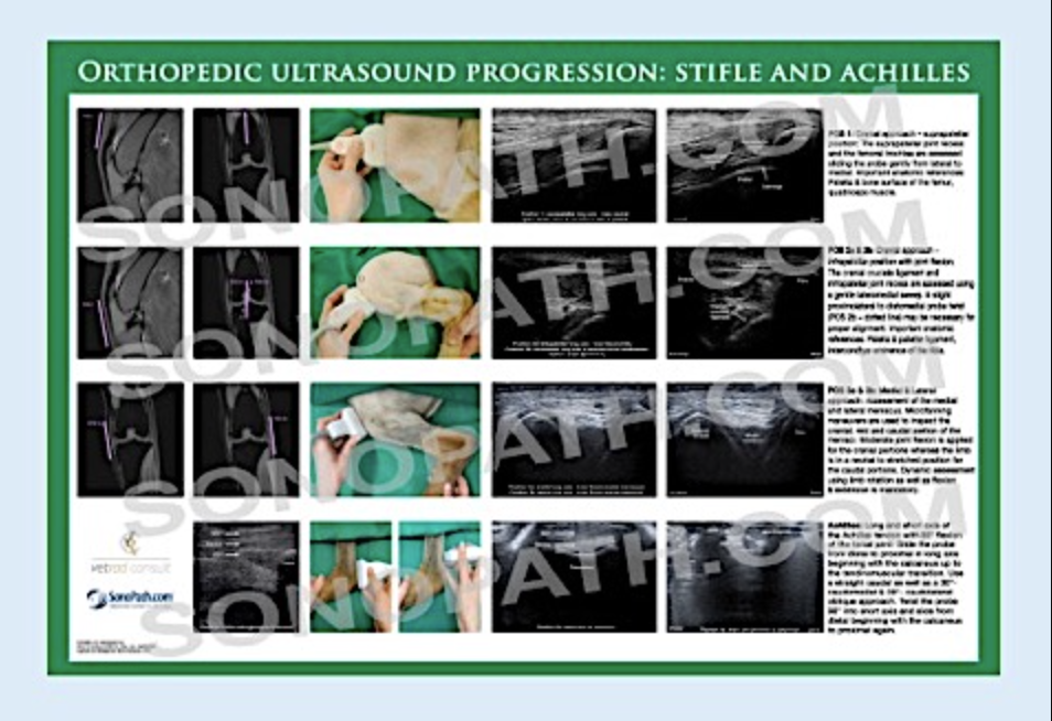
APPLICATION & IMPLEMENTATION OF MUSCULOSKELETAL ULTRASOUND
In the hands of a skilled sonographer ultrasound is an excellent and non-invasive approach to answer common clinical questions in orthopedic patients patients quickly and reliably. The great advantage over cross sectional imaging technique such as MRI, is that ultrasonographic examinations usually do not require any kind of sedation or anesthesia and are associated with significantly lower costs. In orthopedic patients lameness located to the stifle and shoulder joint are the most common indications for ultrasound.
Typical questions in daily practice include:
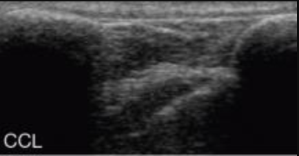
- suspected cranial cruciate pathology with a negative drawer test
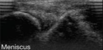
- medial meniscal tears
- recurrent lameness or wound infection after stifle joint surgery
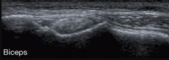
- biceps tendinopathy
- supraspinatus impingement
- fibrosis/contracture of the infraspinatus muscle
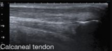
- partial/complete tendon ruptures and monitoring of tendon healing
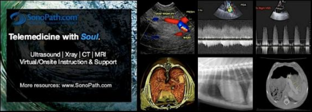
Other common applications are suspected pathology of the brachial plexus and finally any kind of soft tissue swelling/mass within the peripheral soft tissues which may be characterized as inflammatory or neoplastic based on typical ultrasonographic findings easily. Moreover ultrasonography is the most accurate tool in detecting foreign bodies within the superficial soft tissues leaving advanced techniques such as CT and MRI way behind. Orthopedic ultrasound is a rapidly available dynamic technique utilizing our multi-angle video clip approach that is cost effective, minimally invasive and can guide precise sampling for cytological analysis and culture of specific lesions. Orthopedic ultrasound can provide proactive assessment of orthopedic procedures (i.e. stifle surgery technique based on partial vs full cruciate rupture or meniscal tear) in order to refine therapy as well as follow the lesion post therapy. There are literally no limits. If of interest you can scan the bones, joints, ligaments and more in any region of the body…
Pick up SonoPath’s Orthopedic Ultrasound Progression poster to hang in your scanning suite.
A great visual guide to aid in your orthopedic scans.