What is SDEP®?
The Sonographic Diagnostic Efficiency Protocol (SDEP®).
Veterinary medicine has historically been plagued with inconsistencies related to the lack of a standardized protocol for veterinary ultrasound.
The Sonographic Diagnostic Efficiency Protocol (SDEP)® solves this problem, it is
Efficient – Structured – Complete – Repeatable
How Do You Learn SDEP®?
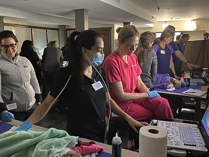
Hands-On Training: 3-Day Weekend labs that consist of wet lab and lectures at our SonoPath veterinary Education center in Andover, NJ.
Live Virtual Ultrasound Training Lab: 3-Day Weekend labs that coincide with hands-on weekends. The virtual attendee’s will have their own dedicated virtual scanning instructor and assistants ready to answer any questions, while you are scanning along from the comfort of your own facility. Click for more details.
Online Courses: SDEP® courses created for you to learn at your own pace. Access to these courses are through our SonoPath Education platform.
Courses are divided into stand-alone modules, designed to deliver more manageable, ‘bite-size’ pieces of information. These courses are a valuable supplement to our hands-on training, and can also be leveraged as a substitute when hands-on training or LIVE virtual is not possible.
Who Can Learn SDEP®?
SDEP® has been taught extensively and successfully to veterinarians and technicians and is widely accepted by interpreting specialists.
How Long Does an SDEP® U/S Take?
STANDARD – typically performed in 10-15 minutes of scan time depending on the sonographer and training.
EMERGENCY – With practice and focus a 5-minute complete abdominal SDEP® protocol is readily achievable and ideal for the complete abdominal evaluation in emergency settings.
The SDEP® protocol is adaptable for speed for emergencies:
Diagnosing trauma scope
Identifies solid organ injury
Ascites
Evaluates for localized and retroperitoneal injury
Heart mass evaluation
Pericardial effusion
Proposed Standardized Procedure for abdominal sonograms in dogs and cats: Sonographic Diagnostic Efficiency Protocol (SDEP®)
Sonographic Diagnostic Efficiency Protocol (SDEP)® was developed by Dr. Eric Lindquist over the course of his 20+ years of extremely busy and intense mobile sonography experience. Mobile sonographers simply cannot afford to miss diagnostic images if they are to maintain a viable mobile ultrasound practice. Mobile sonographers must be extremely efficient in their workflow while continuing to ensure image optimization while scanning. Performing complete sonograms from deep pelvic urethra to gastroesophageal inlet and everything in between, no matter what the animal presentation, provides a complete set of still and video diagnostic images. The SDEP® concepts are also the same for Cardiac SDEP®.
Dr. Lindquist’s evolution of clinical diagnostic sonography workflow, with input from top sonographers worldwide in his community, has amalgamized this clinical workflow into SDEP®. This ensures the sonographer, whether seasoned or novice, does not miss lesions or views (whether abnormal or not) to create optimal and consistent sonograms. Whether the sonographer doing the scan recognizes the structures or not, the sonographer will be able to generate the images necessary for complete interpretation of all structures in the respective cavity, whether abdominal, cardiac, thyroid, eye, or orthopedic SDEP protocols.
Intrinsic image redundancy is ever present throughout the SDEP® scan. This ensures that the interpreter has multiple views of all structures and organs during a direct scan or remotely through telemedicine. The unique scanning protocol includes more challenging views such as the pelvic urethra, pylorus, portal hepatic hylus, both adrenal glands, and the gastroesophageal inlet. The SDEP® scan, as a video clip-based protocol, allows for 3000+ still image equivalent evaluation of the patient as opposed to 30-50 still images protocols of the past with perhaps a few videos.
SDEP® was developed to streamline the sonographer’s learning curve, imaging consistency, and thorough enhanced exam with an endpoint of obtaining an optimized image set for direct and remote interpretation. This begets a better sonographic experience from beginning to end with more consistent and repeatable findings in all animal body types for abdominal and cardiac presentations. SDEP® also provides complete and consistent images and workflow for the interpreter which provides complete and consistent diagnostic interpretation and reporting. SDEP® allows for the best diagnostic care for the patient which is what we all strive to achieve that is repeatable and of the image set quality with which our professional names can be proudly associated.
How do you refine SDEP® once you have completed a SDEP® course?
1 on 1 Enriched SDEP® Remote Training
(special criteria necessary)Refinement of skills offered in hourly segments (1-4 hour increments), with you scanning your animal with a personal instructor via zoom. During these sessions, your instructor can see your hand positions and your ultrasound images on your machine. Designed for those who are SDEP clients and have already taken either the 3-day SEC on-site training or the live virtual training, and need specific refreshers in areas such as adrenals, pelvic urethra, depth, liver etc. Plug and play double camera setup is provided by SonoPath, and sessions are scheduled by appointment.
Our SDEP® Training
What is SDEP® Abdomen?
SDEP® is a 3-second cine loop approach in a 17-point protocol that produces a full abdominal scan, from the deep pelvic urethra to diaphragm and everything in between.
Image shunts
Bile duct obstruction
Renal and bladder pathology
Gastrointestinal obstruction
Pancreatitis, masses
Peritonitis
Mucocele
Ascites
Pericardial effusion
Built-in redundancy of image angles ensures:
Multiple views of every organ and organ system
Minimizes the potential for missed lesions.
Each step is numbered and referenceable, reinforcing communication between instructor, sonographer and specialist.
The sonogram is performed consistently and repeatedly in both traditional scanning scenarios and emergency situations.
Manual probe-hand manipulation avoids probe pressure discomfort and optimizes the acoustic window by minimizing the probe-organ distance and compressing tissue for image consistency.
What is SDEP® Echo?
SDEP® is a 3-second cine loop approach in a 7-point protocol to optimize and evaluate every region of the heart from acquired to congenital disease.
Evaluate functions
Velocities
Pressures
Chamber sizes
3 separate measurements for left atrial
Includes all standard Doppler velocities
Ensures visualization of heart base masses through alternative views of right atrium and auricle
Each step is numbered and referenceable, reinforcing communication between instructor, sonographer and specialist.
The sonogram is performed consistently and repeatedly in both traditional scanning scenarios and emergency situations.