
A shoutout to Dr. Cox at Countryside Animal Clinic for her ingenuity in diagnosing the bladder rupture in this patient and for her excellent case management. Thank you to Sara Hansen SDEP® Certified Clinical Sonographer of Animal Sounds NW for providing the diagnostic images that aided in the diagnosis in this patient. Lastly, thank you to SonoPath’s Specialist, R. McKenzie Daniel, DVM, DABVP for his comprehensive report and recommendations.
PRESENTING CLINICAL SIGNS
Presenting complaint: Vomiting clear liquid for 3 days. Two-day duration of anorexia. Decreased water intake. Has not used litterbox.

Abnormal PE:
T – 95.4
P – 148
R – 24
Tacky mucous membranes, sunken eyes, distended abdomen, generalized muscle atrophy, lethargy, weight loss, hypothermia.
Current Medications: Advantage applied 1/2/25
Labwork Findings:


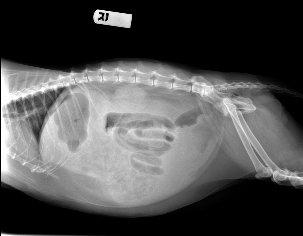

Bubble study using agitated saline injected into urinary catheter to document bladder rupture

ULTRASONOGRAPHIC EXAMINATION OF THE ABDOMEN
Urinary System
The urinary bladder initially presented collapsed in size. Catheterization with injection of agitated saline revealed agitated saline within the urinary bladder lumen as well as outside the urinary bladder within significant primarily anechoic peritoneal effusion. A moderately extensive irregular nonhomogeneous mass occupying the majority of the cranial bladder was present measuring ~3.0 x 1.8 cm. Overtly normal proximal urethra structure and tone were noted to a depth of 2.0 cm.
No evidence of pathology in the area of the aortic trifurcation.
Normal size and margination were present in the kidneys. A normal 1:3 cortex / medulla ratio was maintained. The medulla and cortices were uniform in texture with some increased echogenicity and mild loss of corticomedullary symmetry and definition expected for the age of the patient. No evidence of pelvic dilation was present. The left kidney measured 4.6 cm in length. The right kidney measured 4.1 cm in length.
Free Abdomen
No overt significant omental lymphadenopathy was visualized. Generalized mild omental hyperechogenicity was noted.
ULTRASONOGRAPHIC FINDINGS
- Ruptured urinary bladder with moderately extensive urinary bladder mass, secondary uroabdomen
- Structurally unremarkable visualized gastrointestinal tract with gastric and segmental intestinal ileus
- Normal volume liver
- Mild chronic renal changes
INTERPRETATION OF THE FINDINGS & FURTHER RECOMMENDATIONS
Correlation with effusion analysis with correlation between effusion and serum creatinine and potassium level is recommended. If patient can be stabilized, exploratory laparotomy with gross inspection of the urinary bladder and gastrointestinal tract with potential resection of urinary bladder mass could be considered. However, pending additional diagnostics, an extremely guarded to unfavorable prognosis is indicated.
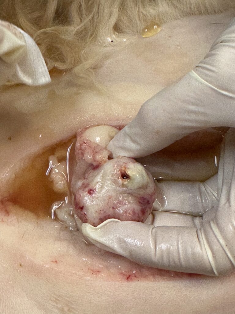
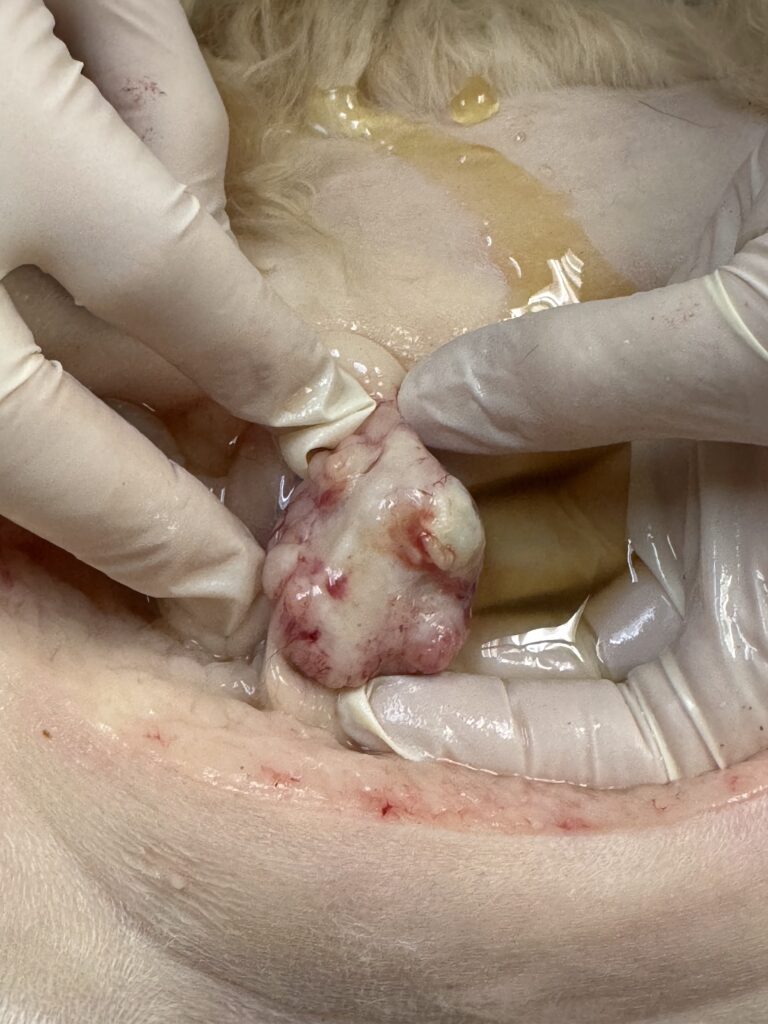
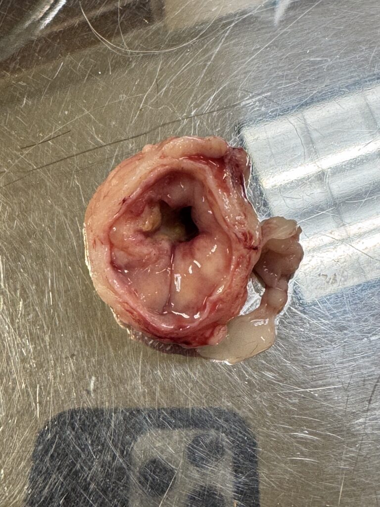
SURGICAL PHOTOS:

Patient Outcome:
This patient did undergo surgery to debulk the tumor at the apex of the bladder wall. However, the patient succumbed to his disease while recovering from surgery. Histopathology was not available.
Discussion:
Bladder tumors are uncommon in the feline patient. Approximately 60% of feline urinary tract tumors are transitional cell carcinoma (TCC). The most common clinical sign is hematuria, but may vary from no clinical signs to stranguria, dysuria, pollakiuria, hematuria, obstruction, anorexia, vomiting, and lethargy. Unlike canine patients that typically develop TCC near the trigone, the feline patient may develop TCC anywhere in the bladder. Diagnosis can be achieved by percutaneous aspiration, but tumor seeding into the body wall is a risk. Neoplastic cells may be evident in urine. In addition, traumatic catheterization can be used to obtain a sample for histopathology.
In this case, the urinary bladder was initially compressed. Based on bloodwork, a post-renal azotemia was suspected. The ascites was suspicious for urine. This prompted a bubble study to be performed. A red rubber catheter was placed in the urethra and agitated saline was injected. In the video sequence, air bubbles can be seen at the apex of the bladder confirming bladder rupture. In addition, a large mass at the bladder apex was visible when the bladder was distended.

Looking to enhance the range of your ultrasound diagnostic efficiency?

Citations:
Burk, Ronald L., and Daniel A. Feeney. Small Animal Radiology and Ultrasonography: A Diagnostic Atlas and Text. Saunders, 2003.
Cristal, Mitchell A., et al. The Feline Patient Editor, Gary D. Norsworthy… (et.Al.). Blackwell Publishing, 2006
Schmidt, Bradley R. “Tumors of the urinary bladder.” Tumors of the Kidney, Bladder, and Related Urinary Structures, Jan. 2004, pp. 241–361, https://doi.org/10.55418/1881041883-3.