A 13-year old, male neutered Pitbull presented to their rDVM with a neck mass. Ultrasonography of the thyroid region was performed by Shari Reffi, CVT with clinical interpretation provided by our very own Eric Lindquist, DVM, DABVP, Cert. IVUSS. The CT portion of this exam was interpreted bv Sebastian Jawinski, German Board Certified Veterinary Specialist in Diagnostic Imaging.
Thank you Dr. Migliaccio at Chester Animal Hospital for your referral and trust in us! Your patient care was exceptional!

History:
UA Results: USG 1.041
CBC:
WBC – 3.9; N 6-17X109/L
LYM – 0.54; N 1.0-4.8X10⁹/L
MONO – 0.18; N 0.2-1.5 X 10⁹/L
NEU – 2.72 ; N 3.0-12.0 X10⁹/L
PLT – 153; N 165-500X 109/L
Chemistry:
T.P. 5.7; N 5.0-7.4g/dL
Glob 3.8; N 1.6-3.6g/dL
Cholesterol – 366; N 92-324/dL
T4 – 1.1; N 0.8-3.5ug/dL
Free T4 by Equilibrium Dialysis. 14.4; N 8-40pmol/L
TSH – 0.47; N 0-0.60 ng/mL
Ultrasound Examination:
The left thyroid lobe was unremarkable, measuring 4.0 mm, with normal parathyroids. The right thyroid lobe revealed an expansive mass with capsular expansion without capsular escape. A moderate amount of blood flow was present. The trachea, regional vessels, and esophagus all appeared intact.
Ultrasound Findings:
Right thyroid mass – appears resectable and encapsulated. Most consistent with thyroid carcinoma.


INTERPRETATION OF THE ULTRASONOGRAPHIC FINDINGS & FURTHER RECOMMENDATIONS :
CT evaluation for surgical planning would be the ideal, or direct surgical intervention.
After the ultrasound results, the patient was referred to our SonoPath Imaging Center for a cervical CT. A cervical CT is recommended as it allows visualization of the tumor and helps to rule out metastasis to regional lymph nodes. In addition, it aids in determination of how invasive the mass is into the surrounding tissues.
The presenting clinical signs stated “Ultrasound shows likely thyroid tumor. Surgical planning.”
COMPUTED TOMOGRAPHIC FINDINGS :
The displayed bony structures of the skull, spine and chest are inconspicuous.
Neck:
There is a rounded to oval-shaped, well-defined thyroid mass noted on the right measuring approximately 4.3 x 2.8 cm, still appearing encapsulated with an heterogenous inner texture and marked contrast uptake. Invasion of the adjacent structures (trachea, carotid artery, jugular vein) is not evident. Some tortuous, hyperplastic vessels are recognized rostral and caudal to the lesion.
The left thyroid gland is unremarkable.
The soft tissues of the head and neck, especially the medial retropharyngeal lymph nodes, are symmetrical and inconspicuous.
Chest:
The displayed parts of the cranial chest including the cranial mediastinum show no particular findings.
COMPUTED TOMOGRAPHIC DIAGNOSIS :
Right thyroid mass currently without overt signs of a locally invasive/aggressive behavior.
INTERPRETATION OF THE FINDINGS & FURTHER RECOMMENDATIONS :
The CT findings of the right thyroid gland are suspicious for a neoplastic process as seen with thyroid adenoma/-adenocarcinoma. Involvement of the adjacent structures is currently not recognized. This does not fully exclude invasion of the supplying vessels. Resection of the right thyroid gland may be curative from a CT perspective.


HISTOPATH:
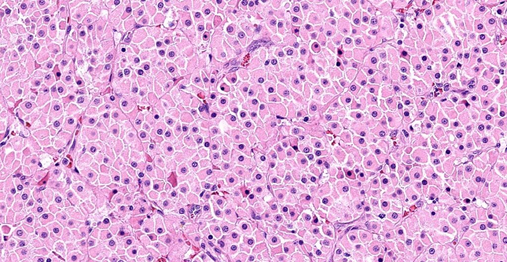
Microscopic Findings: THYROID FOLLICULAR CARCINOMA
Margins: Narrowly excised; Less than 0.1 cm circumferentially.
Angiolymphatic invasion: Not observed.
Microscopic Description:
The right thyroid mass is characterized by solid sheets, clusters and occasional follicle-like formations. The structures consist of diffusely mildly pleomorphic cuboidal to polygonal-shaped epithelial cells. Mild anisokaryosis of round oval nuclei is observed. Delicate fibrovascular stromal bands bisect throughout the mass. There are also some areas where the stroma is more dense. The mitotic count (the number of mitoses counted in 10 HPF 2.37mm2) is 18. Some follicle formation is observed within the mass. There are also some areas of hemorrhage within the mass. The mass is not apparently infiltrating through the surrounding thyroid capsule. Definitive vascular invasion is not observed.
Patient outcome:
The patient underwent surgery and a thyroid follicular carcinoma was removed with narrow margins and no angiolymphatic invasion. Patient recovered well from surgery and has had no further issues to date.
SIGNIFICANCE OF THYROID TUMORS :
Thyroid tumors make up approximately 1-3% of all tumors in the dog with a breed predisposition in Boxers, Golden Retrievers, Beagles, and Siberian huskies. Generally, they occur in dogs aged 10 years or older. Most thyroid tumors are found to be carcinomas and adenocarcinomas. Adenomas account for less than 10% of all thyroid tumors. Most often, they are found by owners that notice a unilateral or bilateral ventral neck mass. However, clinical signs can be attributed to tumor compression on adjacent structures: esophagus, trachea, cervical musculature, and recurrent laryngeal nerve. In addition, those with functional thyroid tumors (less than 15%) may display signs of hyperthyroidism: weight loss, tachycardia, restlessness, and panting. In this case, the T4, Free T4, and TSH suggest a non-functional tumor, which is by far more common. Approximately 60% of dogs have normal thyroid function, while 30% are hypothyroid due to destruction of the thyroid gland by neoplastic tissue, and only 10% show signs of hyperthyroidism. Carcinoma accounts for 85% of thyroid neoplasia.
Ultrasonography plays an integral part of examining this “small part” by allowing confirmation of the tumor being confined to the thyroid. In addition, ultrasound guided FNA may allow the clinician to achieve a diagnosis. Treatment options then are surgery, chemotherapy, radioactive iodine, radiation therapy, and antithyroid drugs such as methimazole. CT, as in this case, is used to determine the surgical resectability of the tumor. Because no invasive/aggressive behavior was noted on CT, the patient was able to be taken to surgery for tumor removal.
FUN FACTS ON SCANNING:
- Occasionally, a thyroid storm or thyrotoxic crisis can occur in animals suffering from a functional thyroid carcinoma.
- Linear probe and microconvex probe both come in handy with this scan.
- Microconvex allows deeper penetration into the tissue of a large thyroid tumor, but will not give the high resolution of a linear probe.
- Make sure to completely shave and use lots of gel for better visualization.
- FNA of thyroid tumors are often hemodiluted due to the vascularity of the tumor.
Looking to enhance your scanning abilities in
ultrasound diagnostic efficiency?

Here are a few of our recommendations:
