

Cooper presented to Conestoga Animal Hospital due to consistent urinary leakage. He was able to urinate intentionally with a good stream but otherwise dribbles urine throughout the day. There was no improvement with Clavamox.
Bloodwork was unremarkable and his urinalysis showed normal concentrating ability with trace protein and 2–3 WBC/hpf. There was no bacteria present. An abdominal radiograph did not show any obvious urinary calculi. An abdominal ultrasound was performed by Dr. Renee Trionfetti showing hydronephrosis, hydroureter, and an ectopic right ureter. Dr. Eric Lindquist reviewed the study and recommended a CT scan to assess renal function and insertion site of the right ureter as well as to plan for surgical correction. The patient was referred for a CT scan and right nephrectomy with Blue Pearl Pet Hospital Wyomissing. Cooper recovered uneventfully and is doing well. The right kidney was sent out for histopathology.
Sonographer: Renee Trionfetti, VMD, 4Paws Veterinary Ultrasound, LLC
Specialist: Eric Lindquist, DMV, DABVP, SonoPath, LLC
Referring Veterinarian: Sarah Urban, DVM, Conestoga AH
Surgeon: Nichole A. Danova, DVM, Blue Pearl Wyomissing, PA
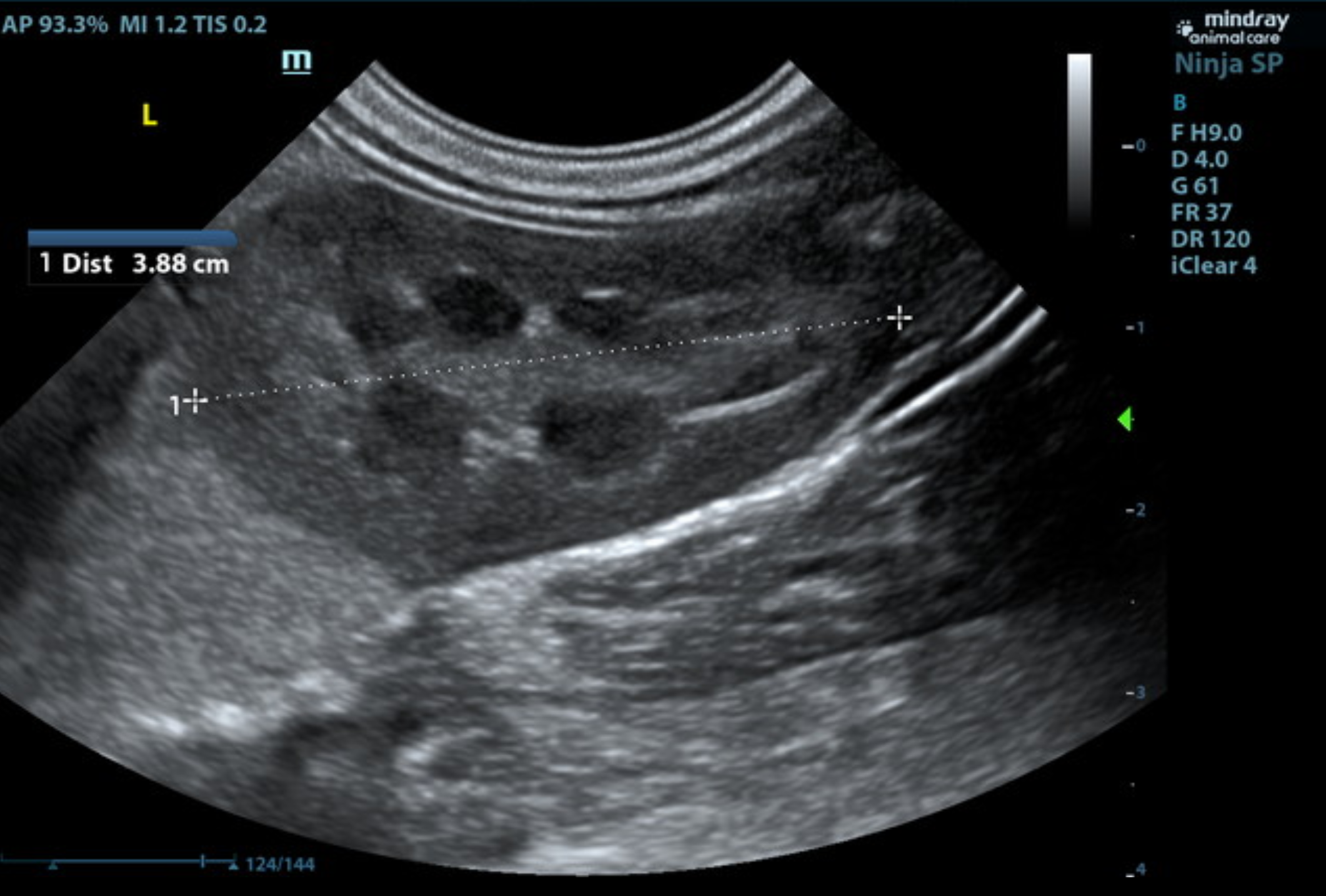
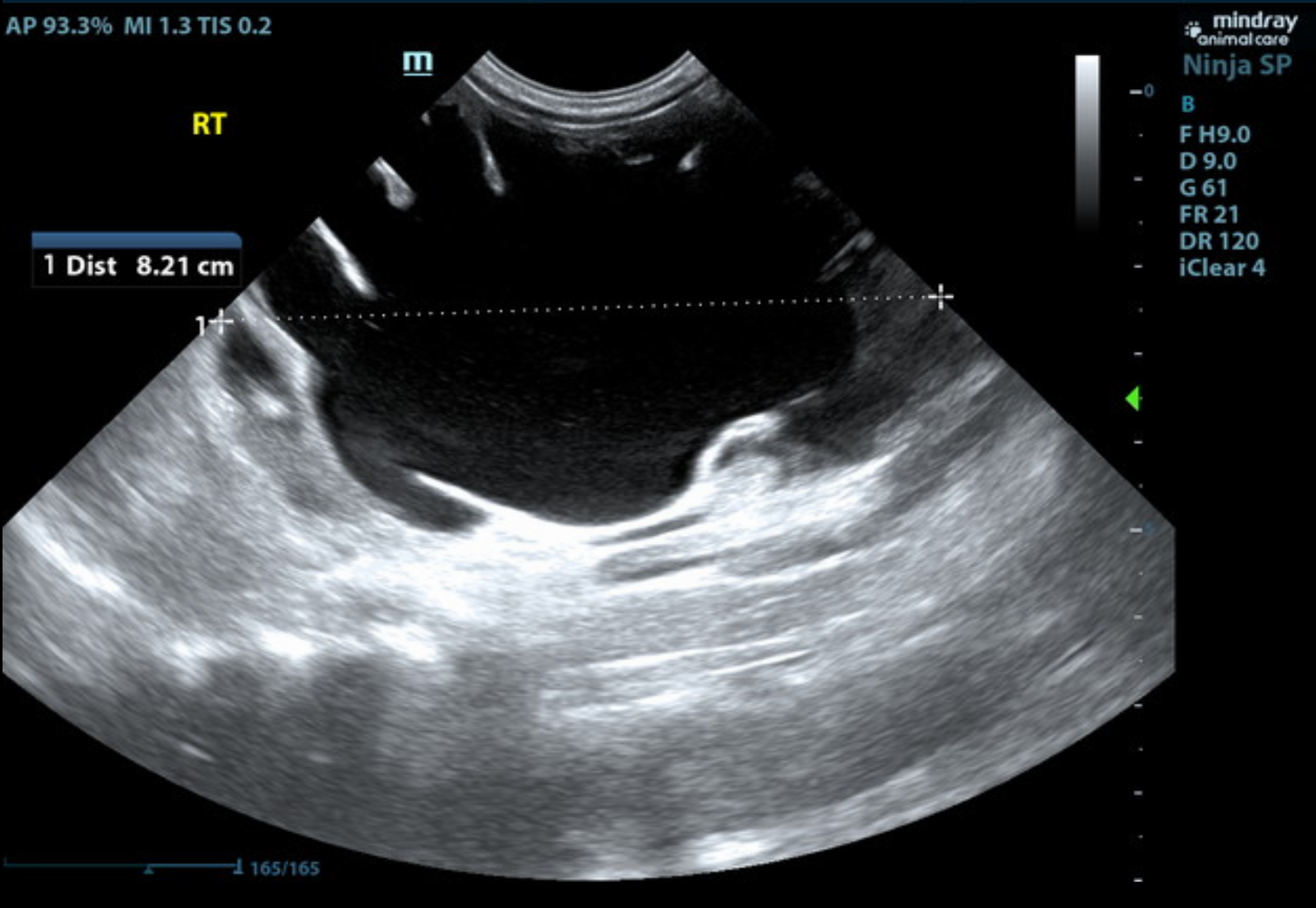
Video 2: US video SDEP® Position 14 showing hydronephrosis of the right kidney
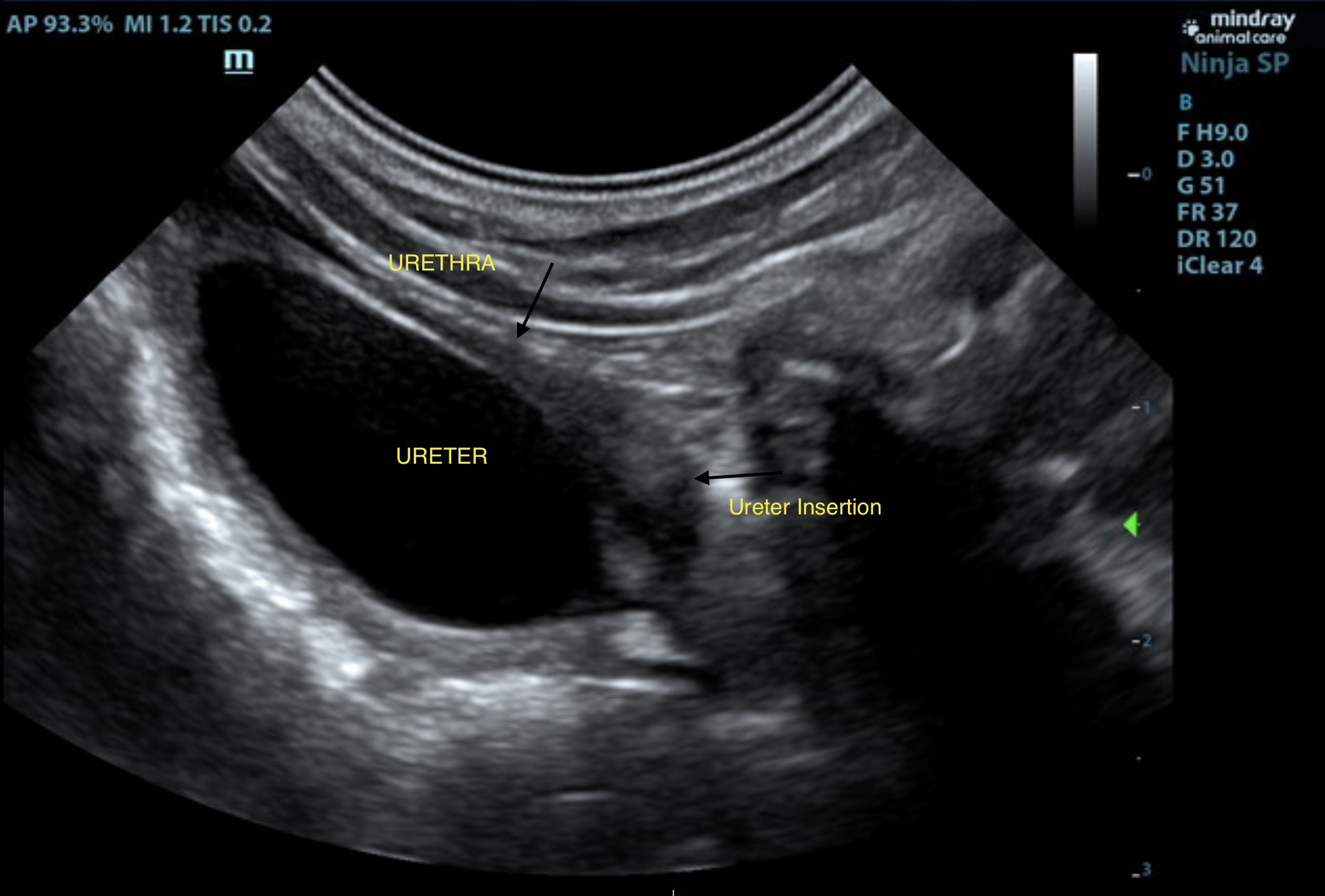
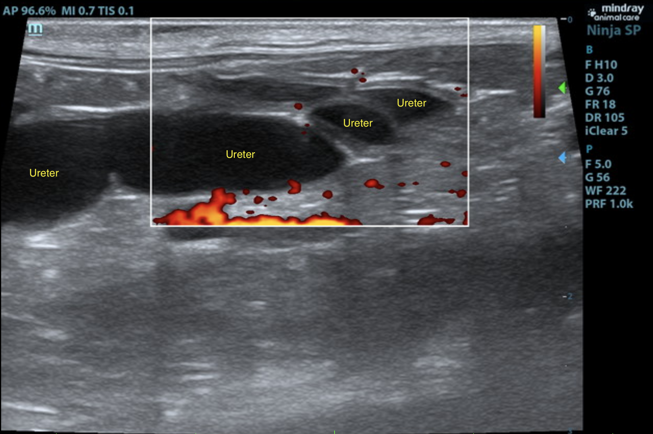
Video 1: US Video at SDEP® Position 1 showing the bladder (most ventral) and the dilated tortuous right ureter dorsal to the bladder
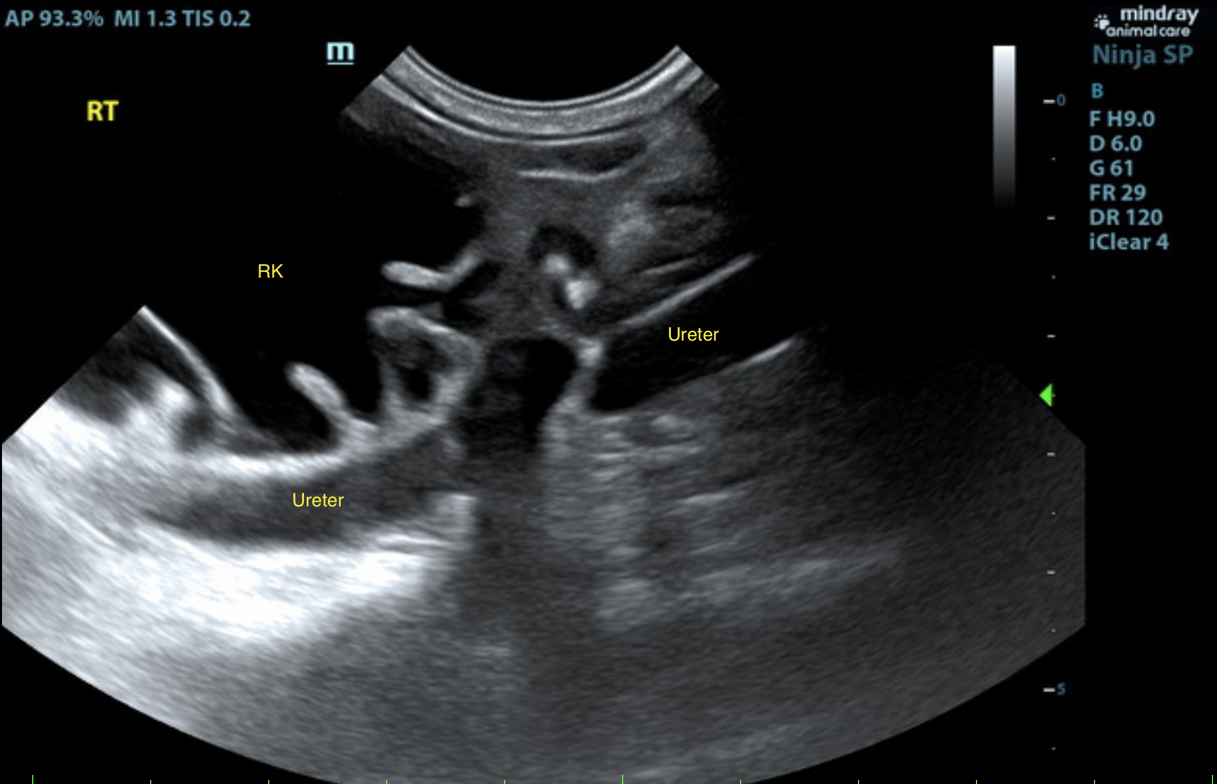
Video 3: US video SDEP® Position 15 showing the dilated right ureter with power Doppler as it courses caudally through the abdomen. The power Doppler confirms this is the ureter and not a vessel.
Urinary System
The urinary bladder presented normal thicknesses and normal tone. No uroliths or sediment were
visualized, and anechoic urine was present. The left kidney presented normal in size, contour, and structure. Left ureter and left kidney appear to be intact and functional.
The right kidney revealed severe hydronephrosis and generalized enlargement measuring 8.2 cm. The right ureter was dilated, bypassing the trigone with maximum dilation measuring 0.85 cm, and appeared to be entering into the distal pelvic urethra approximately 2.0 cm past the cystourethral junction. The ectopic ureter was power Doppler negative, confirming ureteral origin.
Adrenal Glands
Both adrenal glands were visualized and recognized as having normal shape, size, position, and echogenicity for this breed. The phrenic vasculature, glandular echogenicity, and detail were unremarkable. Capsule, cortex, and medullary definition were normal for this age patient. The left adrenal gland measured 1.25 cm x 0.22 cm width at the cranial pole and 0.26 cm width at the caudal pole. The right adrenal gland measured 1.07 cm x 0.35 cm width at the cranial pole and 0.26 cm width at the caudal pole.
Spleen
The spleen presented a smooth, homogeneous parenchyma hyperechoic to liver and renal cortical parenchyma. The capsule was smooth without noticeable expansion or deviation from within the spleen or adjacent pathology. The splenic vasculature demonstrated normal volume without signs of congestion or thrombosis. No sonographic evidence of acute or chronic inflammatory, neoplastic, or infarctual changes were noted.
Liver
The liver images submitted revealed subjectively normal liver size, contour, and structure.
Parenchymal echogenicity was naturally coarse and hypoechoic to the spleen. Vascular and biliary tracts were of normal volume with no evidence of congestion. The gallbladder presented acceptably thin walls with primarily anechoic content. The cystic and common bile ducts were normal. No pathological hepatic lymphadenopathy was evident. No overt structural evidence of inflammatory, infiltrative or regenerative pathology was evident.
Gastrointestinal
Examination of the gastrointestinal tract revealed a stomach and intestine free of stasis, with normal wall thickness, acceptable curvilinear mural detail, and peristaltic activity. Small and large intestine demonstrated normal luminal chyme and stool consistency respectively. No obstructive or overt infiltrative disease was noted. No associated abnormal lymphatic activity was noted.
Pancreas
The base and limbs of the pancreas were observed to be largely isoechoic to surrounding omental fat. Pancreatic duct and capsular contour were acceptably normal and parenchyma respected normal curvilinear patterns. No overt evidence of active inflammatory or neoplastic disease was noted.
• Right ectopic ureter, hydroureter and severe hydronephrosis.
It is doubtful that the right kidney is of functional value in this patient. Although this would be based on contrast CT, minimal visible cortical parenchyma is present. Right nephrectomy/ureterectomy may be the best option in this patient. However, this would also be based on CT evaluation and contrast. Surgical consult is warranted based on CT and ultrasound results.
An ectopic ureter is a congenital abnormality in which one or both ureters terminate in an abnormal location that is usually distal to the bladder trigone.
Ectopic ureters can be intramural or extramural. Intramural ectopic ureters enter into the bladder wall and tunnel through the bladder submucosa inserting into the urethra distal to the urethral sphincter. Extramural ectopic ureters do not enter the bladder wall but rather insert directly into the urethra distal to the urethral sphincter. The majority of ectopic ureters in dogs are intramural.
Patients usually present with a history of continuous or intermittent urinary incontinence. Male patients usually show less signs of incontinence when compared to females. This may be due to increased pressure within the external urethral sphincter as well as the increased length of the urethra in males. Patients with secondary hydronephrosis may also present with a distended abdomen, mass effect on palpation, or poor body condition. Urine scalding and urinary tract infections are common with these patients.
Ultrasound, CT scan with contrast, and cystoscopy are commonly used tools to identify ectopic ureters and hydronephrosis. Surgery is the treatment of choice for ectopic ureters. Cystoscopic guided laser ablation can be used in both males and females. Laser ablation is used to ablate tissues that form the medial ectopic ureteral wall and allow for repositioning of the ureteral orifice into the urinary bladder. In cases of hydronephrosis, nephrectomy is the treatment of choice.
Persistent urinary incontinence is the most common complication following surgical correction. Several studies have shown that approximately 75% of dogs with ectopic ureters who undergo surgical correction achieve continence. Adjuvant medical therapy can help in cases that remain incontinent. 1 2 3
Computed Tomography—Abdomen
There is severe hydronephrotic enlargement of the right kidney and hydroureter of the right ureter. The right hydroureter takes a tortuous course as it is followed caudally and has an abnormal insertion at the bladder where it tunnels caudally dorsal to the urethra and becomes even more dilated before tapering/merging with the urethra within the pelvic canal. The left ureter appears within normal limits in size and insertion at the bladder trigone, resulting in normal passage of contrast medium into the dependent bladder lumen on the later scans, and the left kidney is normal in size (4.2cm) and enhancement without evidence of pelvic dilation.
Assessment
Hydronephrosis and hydroureter – severe, right sided, secondary to a partially obstructed right-sided ectopic ureter with tunneling. No evidence of left sided ectopic ureter or hydronephrosis or other abnormalities. Consultation with a surgery service and/or ideally a service that performs cystoscopic laser ablation is recommended.
Surgery
A right nephrectomy was performed and the ureter was traced to its insertion point and ligated and transected.
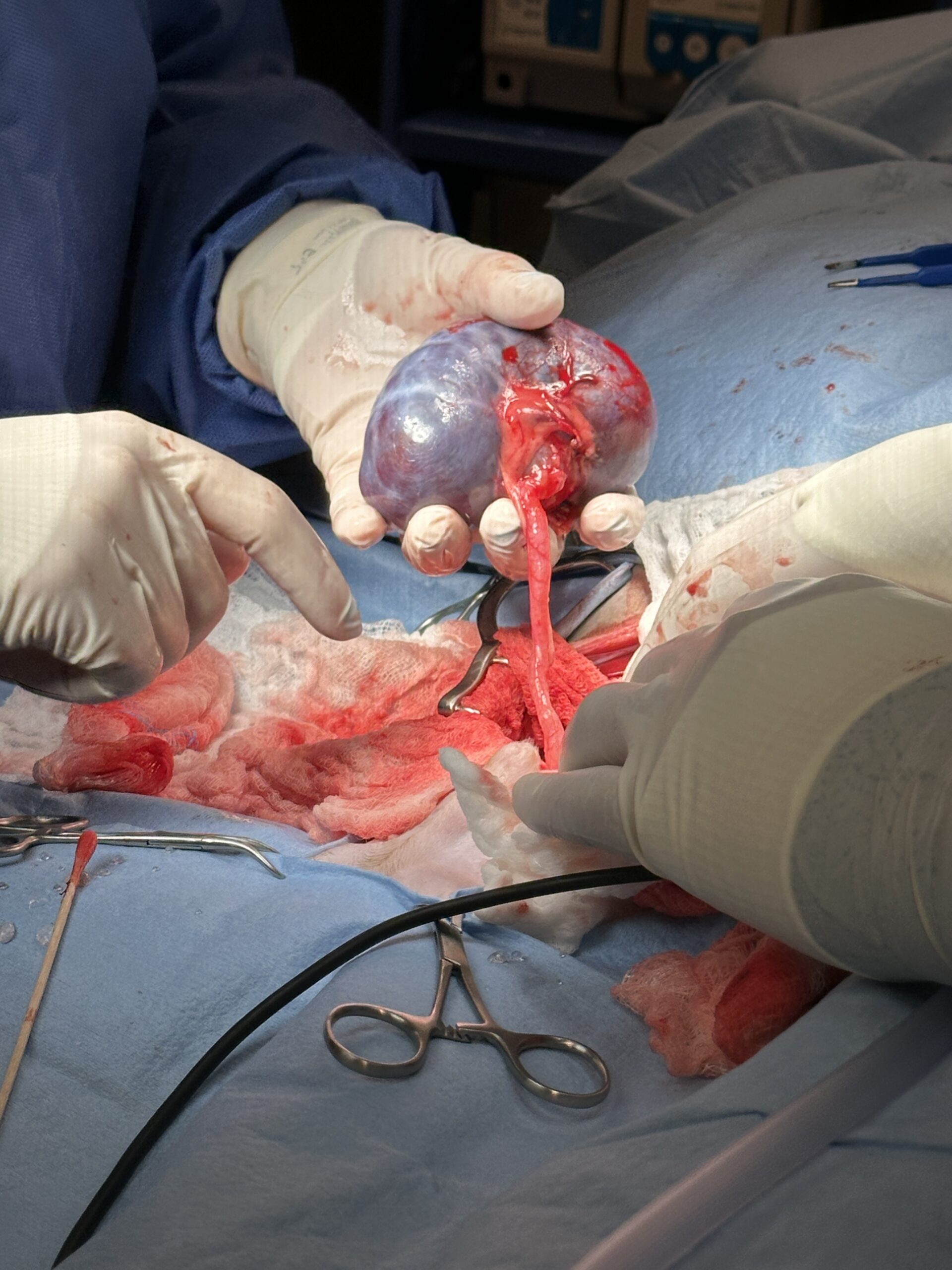
Image showing the hydronephrosis of the right kidney.
Image showing the severe hydroureter.
MICROSCOPIC FINDINGS:
Hydronephrosis, severe, chronic, diffuse
COMMENTS:
This kidney contains histologic changes of severe hydronephrosis, leading to significant renal cortical compression. No evidence of neoplasia or an infectious process is present in the examined sections.
MICROSCOPIC DESCRIPTION:
Kidney: The pelvis is markedly dilated and cystic, compressing the renal parenchyma against the capsule, forming a <1mm rim of variably atrophic renal cortical tissue in some areas. Residual renal tissue contains areas of congestion, edema, and moderate numbers of lymphocytes and plasma cells.
Cooper returned for his recheck exam and suture removal. He is doing very well. There is no incontinence, and he is back to his normal behavior. Cooper’s bloodwork and urinalysis will be monitored now that he only has one kidney.

Intermed/Adv. SDEP® Abdomen
October 8–10, 2026
20 CE credits
Ready for your next-step in ultrasound training? This new lab from SonoPath emphasizes the transition from “How do I get the image?” to “What does this mean clinically?” Through intensive lectures, pathology discussions, and hands-on scanning, participants will strengthen pattern recognition, refine image optimization, and develop a more advanced diagnostic mindset. We can’t wait to see you there!
Sonographic Approach to Urology
1 RACE-approved CE credit ($79.00)
This course provides a systematic approach to imaging one of the most frequently evaluated organ systems in small animal patients, with emphasis on recognizing normal anatomy, identifying pathology, and integrating sonographic findings into patient management.
Check out the Curbside Guide chapter “Urinary” (pg 74–128) and scan the QR Codes to watch video case studies.

References
1. Noël SM, Claeys S, Hamaide AJ. Surgical management of ectopic ureters in dogs: Clinical outcome and prognostic factors for long-term continence. Vet Surg 2017;46:631–641.
2. Reichler IM, Eckrich Specker C, Hubler M, et al. Ectopic Ureters in Dogs: Clinical Features, Surgical Techniques and Outcome. Veterinary Surgery 2012;41:515–522.
3. Gremillion C, Cohen EB, Vaden S, et al. Optimization of ultrasonographic ureteral jet detection and normal ureteral jet morphology in dogs. Vet Radiol Ultrasound 2021;62:583–590.