Did you know that Sonopath has a branch in Canada? Our branch is called The Focal Zone, Canada; this case of the month highlights The Focal Zone, SDEP® Certified Sonographer Crystal Hill, RVT and her ability to capture high end diagnostic views. The case was interpreted by SonoPath specialist R. McKenzie Daniel, DVM, DABVP.
Many thanks to Dr. Debbie Beech from Beech Mobile Pet Services for your referral and trust in us and for your excellent management of this case.

Presenting Clinical Signs/History:
- FELV/FIV negative
- 3 day duration at the rescue
- OVH is planned
Chemistry:
- T.P. – 58.1 (60-80 g/L)
- Creatine Kinase 1358 (25—336 U/L)
- Glucose 9.9 (3.0-8.1 mmol/L)
CBC:
- Lymphocytes 0.54 (1.15-7.5 X10^9/L)
Urinalysis:
- WBC 0-2/hpf
- TNTC cocci
Cefovecin injection administered
PE:
- Palpation of large left-sided abdominal mass – R/O kidney vs other
Ultrasonographic Abdominal Findings:
- Large mid to caudal abdomen thin-walled cystic structure containing primarily anechoic fluid and measuring approximately 6.5 to 7.0 cm in diameter. No evidence of peripheral inflammation.
- Intact left kidney and subjectively intact right kidney.
- Normal urinary bladder.


INTERPRETATION OF THE FINDINGS & FURTHER RECOMMENDATIONS :
Considerations for the intra-abdominal cystic structure may include large omental cyst or significant cystic lymphadenopathy, large cystic ovary, non-obvious significant ectopic hydronephrosis or less likely non-obvious right kidney hydronephrosis or other. The cystic structure is subjectively benign in appearance. US-guided centesis of the cystic structure for fluid analysis cytology +/- C/S can be done, although no overt evidence of inflammatory or abscess criteria could be considered. Abdominal CT may be considered for further clarification and surgical planning given potential location adjacent to major organ vasculature. Otherwise, exploratory laparotomy with gross inspection and potential resection during OVH is warranted.
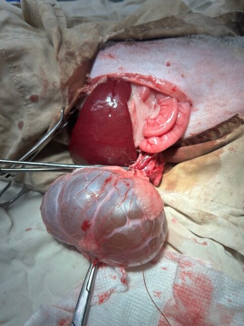
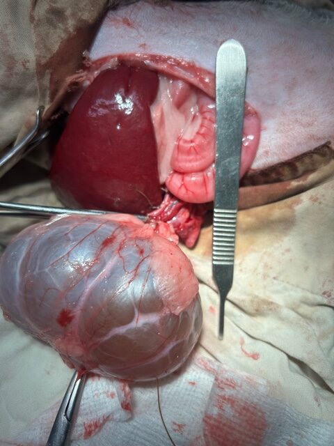
Case Outcome: Based on the ultrasound findings, the patient was taken to surgery where a large ovarian cyst was removed. We are happy to report that the patient recovered without incident and is doing well.


While we do not know what type of cyst was removed from the patient, follicular cysts tend to be the most frequent in the queen. Because these cysts are lined with estrogen-producing cells, persistent estrus, cystic endometrial hyperplasia, infertility, and endometritis may be seen. In addition, these cysts can be partially luteinized causing the excretion of progesterone as well.
Despite many cysts being functional, a significant number are nonfunctional and incidental findings can occur, such as in this case where a mass effect was noted on routine palpation. Ultrasonography can be used to rule out other causes of intra-abdominal masses. And as in this case, ovariectomy or ovariohysterectomy is the treatment of choice.
Looking to enhance your scanning abilities in ultrasound diagnostic efficiency?
Here are a few of our recommendations:
