
in an 11-year-old Female Spayed Pit Bull Mix
This ongoing, complex case is a great example of the use and value of SonoPath’s Internal Medicine Consultation service. The case presented with several layers—beginning with an exam and evaluation by the clinic DVM, followed by an ultrasound submitted to SonoPath for a specialist report. Given the range for potential etiologies and need for a specific, likely long-term management plan, the decision was made to move forward with an Internal Medicine Consultation.
Note that thorough diagnostics were executed on the day of presentation, laying the groundwork for a logical, informed blueprint going forward. No sitting around for this dog!
This patient had a several-month history of intermittent vomiting, decreased appetite, and weight loss when initially presented. Initial assessment revealed non-regenerative anemia and hypoproteinemia, which was attributed to carprofen therapy. She was treated for suspected induced gastric ulceration with omeprazole and sucralfate, and carprofen was discontinued.
Following the initial ultrasound, prednisone was started for suspected inflammatory bowel disease, however, this resulted in a drop in hematocrit. The prednisone dose was reduced, and treatment with omeprazole and sucralfate was restarted. She continued with diarrhea and weight loss. At this point, Dr Geary’s “right arm” technician Keisha Smitley suggested an Internal Medicine Consultation. Given that these consultations had been helpful in previous difficult, nonresponsive cases, the team agreed it was time to bring in another level of expertise.
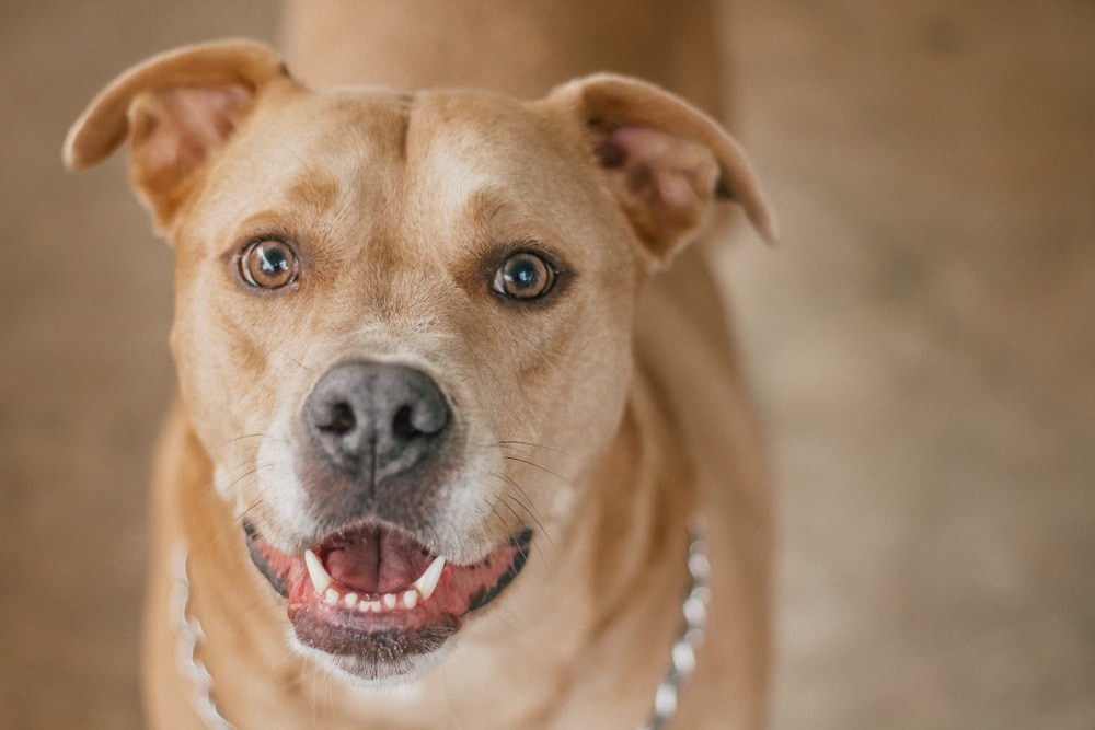
Sonographer: Keisha Smitley, CVT, Geary Veterinary Services, a dedicated, self-driven and experienced technician sonographer who received her ultrasound training from SonoPath
rDVM: Dr. Curtis Geary, owner of Geary Veterinary Services, who lives by the credos “if it’s sick it needs a probe” and full diagnostics on day 1. Dr. Geary is a huge proponent of technicians working at the upper levels of their abilities, within their scope of practice.
SonoPath Specialist: Eric Lindquist, DMV, DABVP, Cert. IVUSS, interpreter of the ultrasound report
SonoPath Specialist: Remo Lobetti, BVSc, MmedVet, PhD, DECVIM, consultant on the internal medicine referral
CBC and Serum Biochemistry 4/1- Non-regenerative anemia and hypoproteinemia
CBC 4/2 and 4/17- No significant abnormalities
Urinalysis 4/2- protein 2+ and USPG 1.042
Serum Biochemistry 4/17- mildly elevatedALT 169 (10-125 U/L) and hyperglobulinemia 5.3 (2.5-4.5 g/dl)
Fecal analysis 4/2– negative for internal parasites
Serology 4/2– negative for vector-borne disease
Abdominal Ultrasound Findings 4/2:
- Splenomegaly with nodules and mass formation
- Small intestinal thickening- strong concern for round cell neoplasia
- Recommendations:
- If the liver is free of evident pathology, proactive splenectomy could be considered; however this would not explain the anemia. Indications for CBC path review, chest radiographs, echocardiogram, and bone marrow biopsy prior to splenectomy.
Survey Radiographs 4/2:
- Moderate interstitial pulmonary opacification and thickening of the bronchial walls consistent with bronchitis
- Normal abdomen
- Multiple sites of chronic arthritis
- Incidental spondylosis
Cytology 4/2:
- Liver- normal cytology
- Spleen – mild extramedullary hematopoiesis and lymphoid hyperplasia
Enlarged spleen with nodule. SDEP® Position 7
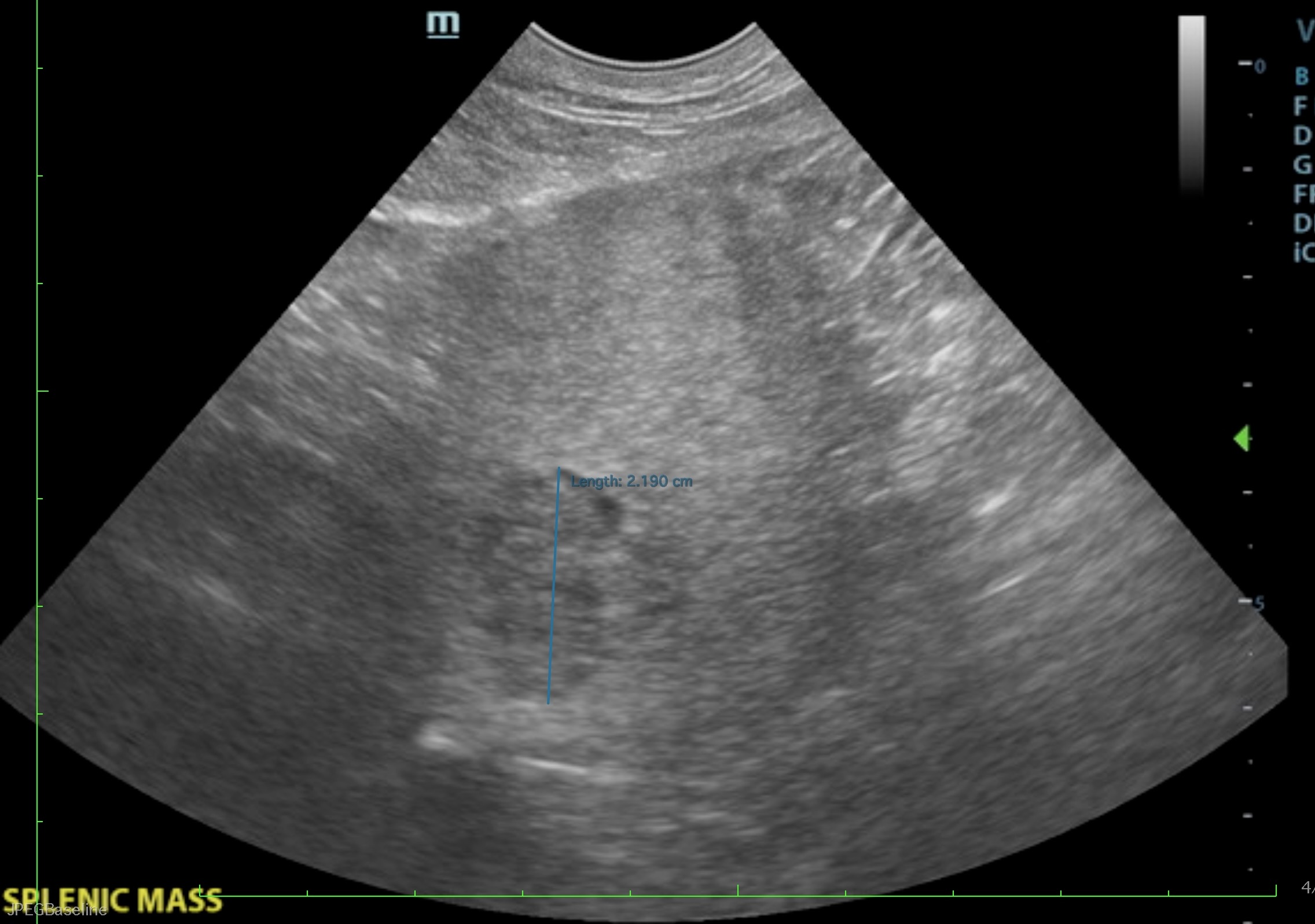
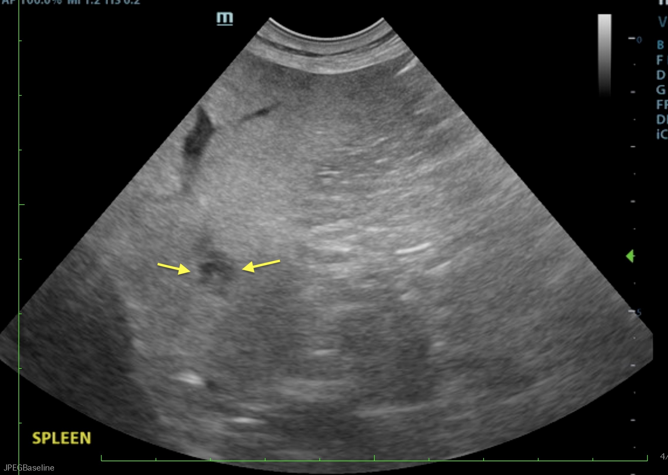
Splenic mass with microconvex probe. SDEP® Position 7
Same splenic mass as in video 2, highlighted using the linear probe. SDEP® Position 7
The history of weight loss, intermittent vomiting, and intestinal thickening seen on ultrasound would be indicative of an underlying enteropathy such as dietary hypersensitivity and inflammatory bowel disease (IBD).
Although splenic cytology was indicative of extramedullary hematopoiesis/reactive hyperplasia, emerging neoplasia should still be considered, especially with the mass.
The mildly elevated ALT activity, hyperglobulinemia, and bronchitis can all be considered incidental findings
The anemia and hypoproteinemia evident on the initial CBC and serum biochemistry is consistent with gastric/duodenal ulceration.
- Serum cobalamin and folate assay
- Endoscopy of the upper GI tract with biopsies
- Splenectomy could be considered, which would also allow for full thickness biopsies of the GI tract
Initial management would involve feeding small, frequent meals of a novel protein/hypoallergenic diet and cobalamin supplementation (if indicated). If there is not a satisfactory improvement, then the addition of prednisolone would be indicated. As there was an adverse reaction to the previous course of prednisone, oral budesonide would, however, be recommended.
Prednisolone forms the foundation of therapy in dogs with IBD, starting at 1 mg/kg BID for 10 days then tapered over 10–12 weeks. Budesonide can be administered as an alternative to prednisolone as it has high topical anti-inflammatory activity and lower systemic activity because of its high affinity to the steroid receptor and rapid hepatic conversion to metabolites with minimal or no steroid activity.
If there is still no improvement or the dose of prednisolone remains high and/or there are unacceptable adverse side effects then considered adding azathioprine (1–2 mg/kg SID for 2 weeks, followed by alternate-day administration) and/or cyclosporine (5 mg/kg SID).
Hypoallergenic/novel protein diet.
Cobalamin deficiency has been associated with GI tract signs despite appropriate therapy in animals with IBD as mucosal repair is impeded. It is given subcutaneously for 6 weeks on a weekly basis, with supplementation continued every 3 weeks for the indefinite future if cobalamin remains low. Oral supplementation with cobalamin is equally effective but must be given daily for 12 weeks before testing and determining the need for ongoing supplementation.
Probiotic administration using either single-strain or multi-strain products can be helpful in the management of inflammatory bowel disease.
Based on Dr. Lobetti’s consultation recommendations, this patient was switched to prednisolone, B12 added, and placed on a hypoallergenic diet—with almost immediate improvement. Over the next several weeks, she was able to be completely weaned off the prednisolone and maintained on the hypoallergenic diet and now monthly B12 injections. Her vomiting and diarrhea have completely resolved and her appetite is normal. She gained all of her weight back, is more active, and wants to play. At this point, the current regimen is working for her. If needed in the future, the management plan outlined by the Internal Medicine Consult can be revisited.
Discover SonoPath’s Educational Telemedicine® and Internal Medicine Teleconsultation Services:
The Curbside Guide (section 3: Gastrointestinal/Pancreatic Pathology, pg 164 on Inflammatory Bowel Disease (IBD) Canine)

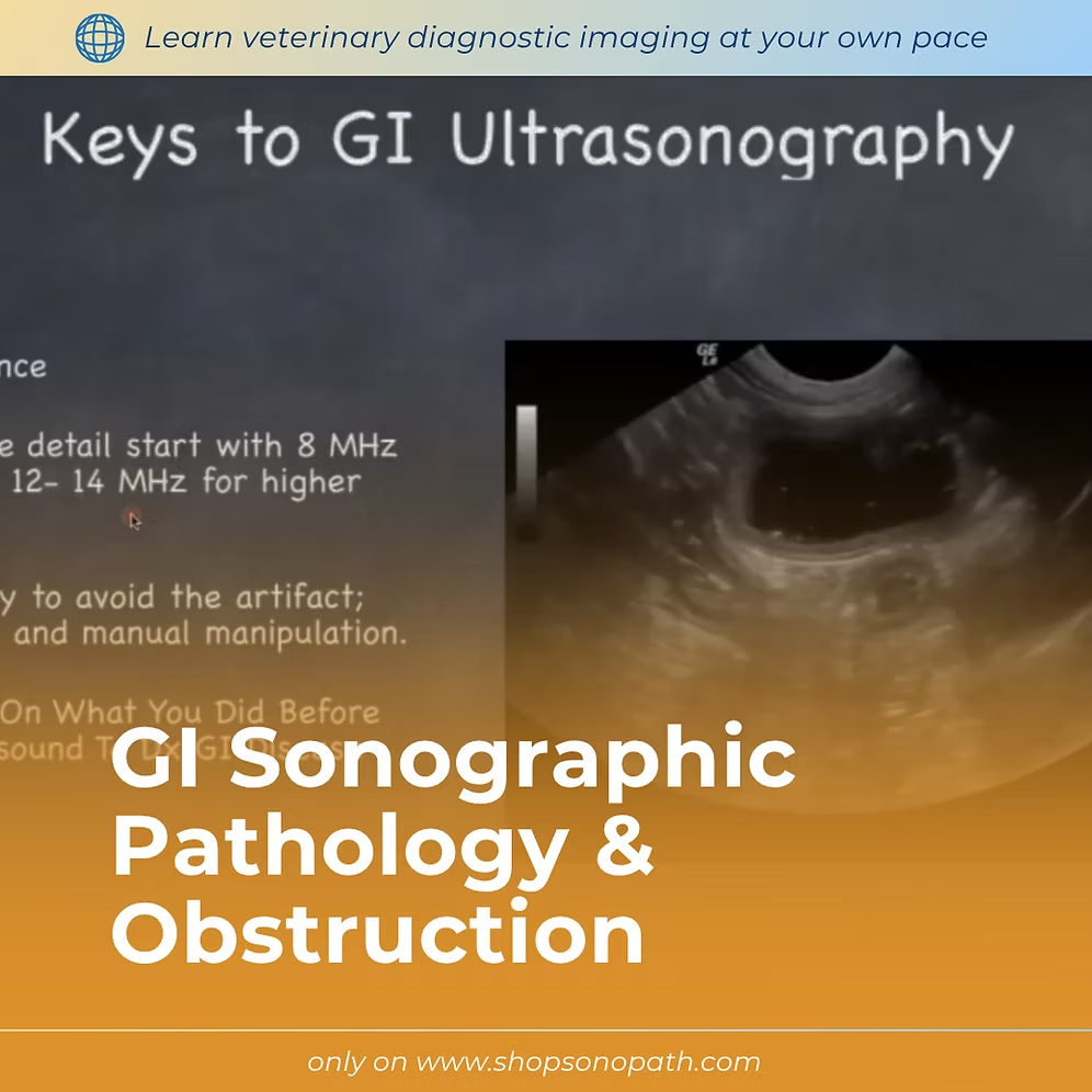
2 RACE-approved CE credits
2 x self-paced video webinars plus post-course CE quiz
Approximate Study Time: 2 hours (self-paced)
Instructor: Eric Lindquist, DMV (Italy), DABVP (Canine & Feline), Cert. IVUSS, Founder & CEO of SonoPath
Develop a stronger understanding of gastrointestinal ultrasound by learning how normal anatomy, function, and disease processes appear from a sonographic perspective. This course reviews gastrointestinal structure, motility, and relationships with surrounding organ systems to help clinicians recognize when findings deviate from normal.
Through practical case examples and pathology discussions, participants will explore common gastrointestinal abnormalities, including obstruction patterns, pyloric disease, and other sonographic presentations that impact clinical decision-making.
The course emphasizes pattern recognition and diagnostic efficiency, helping clinicians understand how changes in gastrointestinal wall structure, layering, motility, and surrounding anatomy can guide differentials and next steps in patient management. A curvilinear approach to interpretation is introduced to improve recognition of abnormal inflammatory, obstructive, and disease-associated patterns.

November 13-14, 2026
16 RACE-approved CE
Designed for advanced veterinary sonographers ready to refine echocardiographic technique, improve diagnostic interpretation, and integrate advanced cardiac pathology into clinical decision-making.
2027 SDEP® Echo Hands-On Ultrasound Training
22 RACE-approved CE credits
Live lectures, hands-on training, & small-group instruction
Designed for veterinarians and veterinary technicians looking to build or advance practical echocardiography skills using a structured, repeatable protocol. Whether you are new to cardiac ultrasound or refining existing skills, the SDEP® Echo Protocol allows personalized training tailored to your experience level. Beginners to echocardiography will build confidence while experienced sonographers refine and develop speed, consistency, and diagnostic accuracy.
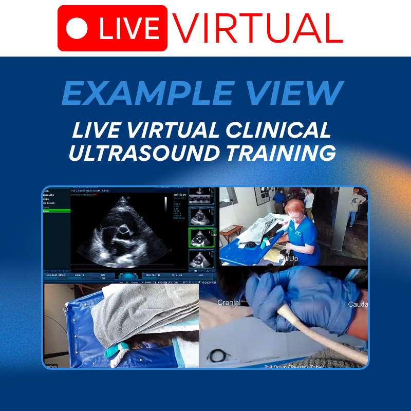
SDEP® Echo REMOTE Live Virtual Training Weekend
22 RACE-approved CE credits
Join us remotely with a direct Zoom link to your SDEP® instructor, you scan from your home machine on your patients training with the watchful support of not only your instructor, but a dedicated breakout room instructor.