Thank you to Dr. Bilinsky for the referral and thoughtful management of our recent feline patient. We’d also like to recognize Queensway Animal Hospital for their ongoing trust and partnership. Our appreciation extends to Crystal Hill, RVT, SDEP® Certified Clinical Sonographer with SonoPath’s Canadian branch – The Focal Zone – for her precise imaging, and to specialist Sara Brethel, DVM DACVIM (Cardiology), for her outstanding interpretation, which offered critical insights for the case. Collaboration like this reminds us of the synergy of a strong, connected team.
History:
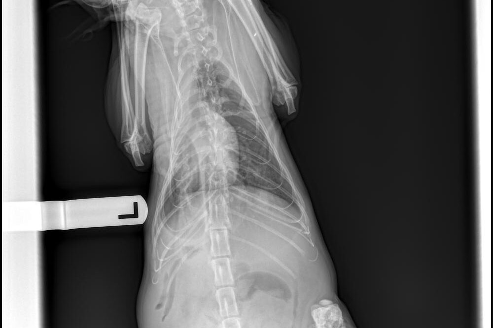
The patient presented for increased abdominal effort. At that time, a pleural effusion was identified and approximately 400 mL of fluid was removed from the thoracic cavity. A prior abdominal effusion had also been documented at the emergency clinic.
Clinical Signs:
Physical examination revealed dull mentation, intermittent inappetence, and increased abdominal effort. Blood pressure today averaged 130/93 mmHg. The patient has been receiving furosemide 20 mg TID, which is currently being tapered while spironolactone is being introduced.
Abnormal Findings (PE/Chem/CBC/UA):
Chemistry: Elevated glucose, SDMA, and BUN
Electrolytes: Hypokalemia, hypochloremia
Hematology: Elevated reticulocytes, WBCs, neutrophils, monocytes, eosinophils, and platelets
ULTRASONOGRAPHIC EXAMINATION OF THE HEART

ECG Interpretation:
There is underlying sinus rhythm with slurring of the ST segment.
Cardiac Presentation:
The left atrium is severely enlarged. The mitral valve leaflets are normal and there is no mitral regurgitation. There is no evidence of systolic anterior motion of the mitral valve and no evidence of a left ventricular outflow tract obstruction. There is concentric hypertrophy of the posterior wall and there appears to be thinning of the interventricular septum. The right atrium is at least mildly to moderately enlarged. The tricuspid valve is normal without evidence of tricuspid regurgitation. The right ventricle appears to have preserved systolic function subjectively. The aortic and pulmonic valves are normal without evidence of insufficiency. Aortic and pulmonic outflow velocities are within normal limits. The aorta and PA are normal along with the associated PA branches. There is evidence of pleural effusion, scant pericardial effusion, and ascites. There is no evidence of an intracardiac mass.


ULTRASONOGRAPHIC FINDINGS
- Hypertrophic cardiomyopathy, ACVIM stage C
- Severe left atrial enlargement
- Mild to moderate right atrial enlargement
- Tricavitary effusion
INTERPRETATION OF THE FINDINGS & FURTHER RECOMMENDATIONS
The slurring of the ST segment on the ECG is due to the severity of the cardiac disease and no specific therapy is indicated at this time.
The patient is at toxic levels of furosemide and this needs to be reduced. Recommend giving 10 mg twice daily. As long as there is no continuation of effusion, then further reduce to 5 mg twice daily.
With the reported hypokalemia, if the potassium is <3, then potassium supplementation is recommended at a dose of 2 ml equivalence per day.
Recommend starting pimobendan at a dose of 0.27 – 0.32 mg/kg twice daily, along with clopidogrel at a dose of 18.75 mg (1/4 of a 75 mg tablet) every 24 hours. Spironolactone can be continued at a dose of 2 mg/kg once daily.
The patient has evidence of left ventricular concentric hypertrophy and is classified as a stage C due to having tricavitary effusion and severe left atrial enlargement. If not already performed, it is recommended to ensure that patients blood pressure is normal and the patient is euthyroid.
The client should start monitoring respiratory rate and effort at home if not already doing so. The resting respiratory rate should be < 35-40 breaths/minute when the patient is resting or sleeping. If the breathing rates are increasing then chest radiographs are recommended.
Recheck fluid assessment and kidney values are recommended in another week, sooner if the patient is decompensating or the kidney values are worsening. If the kidney values are normal and the patient is doing well, recommend starting an ace inhibitor (enalapril versus benazepril) at a dose of 0.5 mg/kg by mouth every 12-24 hours. 2-3 weeks after starting ace inhibition, recheck blood work is advised, specifically kidney values.
With the severity of the heart disease and the severity of the effusion, the patient has a poor to guarded prognosis. Unfortunately, median survival times for congestive heart failure with hypertrophic cardiomyopathy are 6-12 months. Some patients can survive longer periods of time.
Recommend a recheck echo in 3-4 months, sooner if the patient is not doing well.
If the patient continues to effuse despite diuretic therapy, then there is concern for another underlying disease process causing the effusion and submission of the fluid for analysis and cytology would be indicated.
Unfortunately, due to the nature of this condition, this patient is at risk for passing away suddenly.
Patient Outcome: The patient remained in the care of the rescue as a long-term foster due to ongoing costs and was not adopted out. He stayed under their care for several more weeks following his ultrasound scan, but unfortunately, his condition declined, and he was humanely euthanized.
Discussion: What is Hypertrophic Cardiomyopathy?
Hypertrophic Cardiomyopathy (HCM) is a disease of the myocardium. It is primarily seen in cats and occurs rarely in the dog. HCM is the most common cardiomyopathy seen in the cat. In all cats HCM can be diagnosed at any age but males tend to develop severe disease at a younger age. The cause of most cases of HCM is unknown however a genetic mutation has been identified in the Maine Coon and Ragdoll breeds. A suspected mutation has been identified in the Sphynx breed. HCM is characterized by a non-dilated hypertrophied left ventricle. It is typically a diagnosis of exclusion and other diseases causing left ventricular hypertrophy must be ruled out. These diseases include hyperthyroidism, systemic hypertension, acromegaly, lymphoma and pseudo thickening due to dehydration.
The phenotype HCM presents in multiple different ways causing diffuse assymetrical or symmetrical thickening of the left ventricle, specifically in the interventricular septum(IVS) or left ventricular free wall(LVFW). The mutation affects the ability of the cardiac muscle to contract and relax causing a thickening of the left ventricular free wall and septum. The thickened muscle has a decreased blood supply which causes muscle fiber necrosis. The damaged muscle is then replaced with fibrotic tissue further affecting the ability of the heart muscle to contract and relax. This increases left atrial pressure and causes left atrial dilation. In severe HCM, pulmonary edema, pericardial effusion, pleural effusion, ascites and aortic thromboembolism can occur. Some cats may die suddenly and others have a subclinical form of the disease and live a normal life. 1,2
There is no known treatment to slow or reverse HCM. Therapy is typically aimed at treating congestive heart failure(CHF) and arterial thromboembolism(ATE). For cats in CHF furosemide, ACE inhibitors, pimobendan and thoracocentesis have been used to extend life. Clopidogrel(Plavix) has been shown to be superior to aspirin in preventing recurrent ATE formation and increasing survival times of HCM cats.
Citations:
1. Kittleson MD, Côté E. The Feline Cardiomyopathies: 2. Hypertrophic cardiomyopathy. J Feline Med Surg 2021;23:1028–1051.
2. Trehiou-Sechi E. Comparative Echocardiographic and Clinical Features of Hypertrophic Cardiomyopathy in 5 Breeds of Cats: A Retrospective Analysis of 344 Cases (2001–2011). Available at: https://onlinelibrary.wiley.com/doi/10.1111/j.1939-1676.2012.00906.x.
3. Hogan DF, Fox PR, Jacob K, et al. Secondary prevention of cardiogenic arterial thromboembolism in the cat: the double-blind, randomized, positive-controlled feline arterial thromboembolism; clopidogrel vs. aspirin trial (FAT CAT). Journal of Veterinary Cardiology 2015;17:S306–S317.

Looking to enhance the range of your ultrasound diagnostic efficiency?
