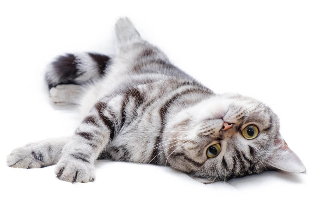
A juvenile male cat was referred to Wilvet South Emergency Hospital, for overnight hospitalization, intravenous (IV) antibiotic therapy, and IV fluid support due to an elevated WBC count identified by the referring veterinarian, lethargy and anorexia. Upon Dr. Natalie Jackson’s examination, the patient was noted to be underweight, a condition reported by the owner to have persisted since early life. A rapid viral screening test (SNAP) performed by the referring veterinarian was negative.
Given the patient’s history of chronic underweight status, failure to thrive, and hyperglobulinemia observed on blood work, FIP was considered a differential diagnosis. The patient’s WBC count was not strongly indicative of infection but was suggestive of systemic inflammation. The patient was afebrile at the time of presentation. A diagnostic ultrasound was recommended, revealing classic medullary rim sign in the kidneys, a finding consistent with FIP. Sonopath specialist Dr. Eric Lindquist (DMV, DABVP, Cert. IVUSS, CEO & Founder of SonoPath) confirmed the suspicion with his detailed abdominal interpretation. The owner declined a feline coronavirus polymerase chain reaction (PCR) test.

Referring DVM labs showed elevated WBC with neutrophilia, elevated total proteins with a low normal ALB and low K and Cl.
Abnormal PE/Chem/CBC/UA Results:
Chem 10: Creatinine 0.6 (low), Total protein >12 (high), ALB 2.2 Electrolytes: Potassium 2.8 (low); Chloride 111 (low).
Thank you Dr. Natalie Jackson for your excellent patient care management, and thank you Dr. Eric Lindquist for your detailed report, confirming the FIP suspicion.
ULTRASONOGRAPHIC EXAMINATION OF THE ABDOMEN
Urinary System:
The urinary bladder, trigone, and pelvic urethra presented normal thicknesses and normal tone to a depth of 3.0 cm. The ureters were not visible which is normal. No uroliths or sediment were visualized and anechoic urine was present. No evidence of inflammatory or neoplastic changes were noted. Ureteral papillae were normal.
The bilateral kidneys in this patient presented uniform corticomedullary ratio; however, a dramatic hyperechoic corticomedullary band was present. This is consistent with bilateral medullary rim sign. Mild degenerative renal changes were noted. The contour was uniform. This is generally an idiopathic finding, yet at times can be related to FIP or lymphoma in cats. Assessment for any proteinuria would be warranted if not already performed. An expansive irregular nodule was noted in the cranial pole of the left kidney with subcapsular halo. The left kidney was hypervascular. The left kidney measured 4.5 cm. The right kidney measured 5.0 cm. A pericapsular inflammatory pattern was noted.
The iliac trifurcation was unremarkable.
Adrenal Glands:
The adrenal glands were uniform, yet bilaterally swollen and hypoechoic. This is most consistent with stress-induced hyperplasia.
Spleen:
The spleen presented a smooth homogeneous parenchyma hyperechoic to liver and renal cortical parenchyma. The capsule was smooth without noticeable expansion or deviation from within the spleen or adjacent pathology. The splenic vasculature demonstrated normal volume without signs of congestion or thrombosis. No sonographic evidence of acute or chronic inflammatory, neoplastic, or infarctual changes were noted.
Liver:
The liver images submitted revealed subjectively normal liver size, contour, and structure. Parenchymal echogenicity was naturally coarse and hypoechoic to the spleen. Vascular and biliary tracts were of normal volume with no evidence of congestion. The gallbladder presented acceptably thin walls with primarily anechoic content. The cystic and common bile ducts were normal. No pathological hepatic lymphadenopathy was evident. No overt structural evidence of inflammatory, infiltrative or regenerative pathology was evident.
Gastrointestinal:
Examination of the gastrointestinal tract revealed a stomach and intestine free of stasis, of normal wall thickness, acceptable curvilinear mural detail, and peristaltic activity. Small and large intestine demonstrated normal luminal chyme and stool consistency respectively. No obstructive or overt infiltrative disease was noted. No associated abnormal lymphatic activity was noted.
Pancreas:
The base and limbs of the pancreas were observed to be largely isoechoic to surrounding omental fat. Pancreatic duct and capsular contour were acceptably normal and parenchyma respected normal curvilinear patterns. No overt evidence of active inflammatory or neoplastic disease was noted.
A mesenteric lymph node cluster was present, the grouping measuring 2.5 cm x 2.0 cm.


(aggressive, major rim sign apparent)

ULTRASONOGRAPHIC FINDINGS:
- Mesenteric lymphadenopathy.
- Aggressive medullary rim kidneys with regional inflammation.
INTERPRETATION OF THE FINDINGS & FURTHER RECOMMENDATIONS:
Strong concern for FIP is warranted. FNA of the lymph nodes and kidneys is recommended for further definition.
Management and Outcome
As the diagnosis preceded the widespread availability of targeted antiviral therapy for FIP, the owner was referred to the FIP Warriors organization in Oregon, an independent group facilitating access to experimental treatment. The patient was subsequently managed under their guidance, with the GS-441524 treatment. Follow-up reports indicated that the patient successfully completed antiviral therapy, achieved a normal body condition score, and underwent routine castration without complications. At the time of the last update, the patient remained clinically healthy and free of FIP-related symptoms.
This case highlights the importance of considering dry FIP in young cats presenting with chronic underweight status and hyperglobulinemia. Diagnostic imaging played a crucial role in supporting the presumptive diagnosis. The patient’s positive response to experimental antiviral treatment underscores the evolving therapeutic landscape for FIP management.
What is FIP?
Feline infections peritonitis is caused by a mutation of the feline enteric coronavirus (FECV). Feline coronavirus is an RNA virus that affects both domestic and wild members of the Felidae family. Approximately 74-100% of cats in shelters, catteries, and multi-cat households are infected with feline enteric coronavirus. Animal are usually asymptomatic for FECV, but it can manifest as a mild diarrhea. In approximately 10% of cats infected with FECV, the virus will mutate into the more virulent form, feline infectious peritonitis. The FIP disease is believed to be due to an immune-mediated response to the viral antigen. FIPV attracts antibodies, macrophages, and neutrophils, which leads to complement fixation, which causes increased vascular permeability. Infection of macrophages lead to the development of granulomatous lesions.
FIP generally is displayed in 2 forms: the effusive, wet form or the granulomatous, dry form. A third “mixed” form has been described. Wet FIP is generally seen 4-6 weeks following after a stressful event, such as spaying/neutering or rehoming. Clinical signs are secondary to a weak cell mediated response causing vasculitis and leakage of protein and fluids into body cavities (generally chest or abdomen). The dry form of FIP is more likely due to a partially successful cell-mediated immune response and generally granuloma formation. Occasionally wet FIP may be a sequelae of the end stage dry form.
Treatment options have traditionally been limited. Owners have been forced to seek treatment with the FIP warriors facebook group. Historically, they have been using the GS-441524 nucleotide for treatment. In addition, both remdesivir (the prodrug of GS-441524) and molnupiravir, have been used for treatment. Both remdesivir and molnupiravir are drugs that have been used to treat human COVID-19.
In this case, the patient was treated alongside FIP warriors with serial monitoring performed. He successfully completed his treatment with GS-441524 with no complications. He was back to a normal body condition at the time of neutering, and he is now living his best life without FIP!
Citation:
- Coggins, Sally J. “FIP in Cats: Feline Infectious Peritonitis.” Vca, 2023, vcahospitals.com/know-your-pet/feline-infectious-peritonitis
- Little, Susan E. The Cat: Clinical Medicine and Management. Elsevier, 2025
- Colleran, Elizabeth. “Feline infectious peritonitis.” AccessScience, 2023, https://doi.org/10.1036/1097-8542.757283.
Looking to enhance the range of your ultrasound diagnostic efficiency?

