
in an 8-month-old Maltese
An 8-month-old, intact, male Maltese presented to Village Veterinary Hospital for a routine exam, brought in from a local Rescue as part of a recent transport. On exam, a 6/6 heart murmur was confirmed and a cardiac ultrasound was immediately scheduled with SonoPath’s New Jersey Mobile team.
Sonographer: Rebecca Hamilton, SDEP® Certified Sonographer with SonoPath’s New Jersey Mobile
Specialist: Bradley Harris, DVM, DACVECC, DACVIM
rDVM: Amanda Kiselak, VMD, Village Vets

Blood Pressure: Performed on the left front with a #2 cuff:
114/82 (93)
133/95 (108)
172/138 (149)
Heartworm negative
Screening bloodwork within normal limits
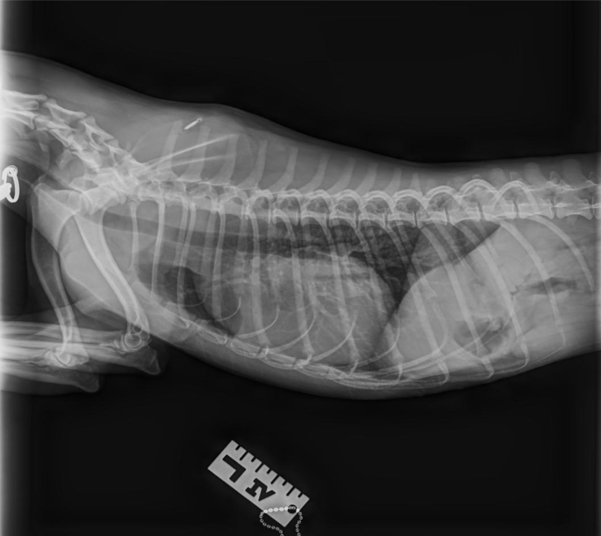
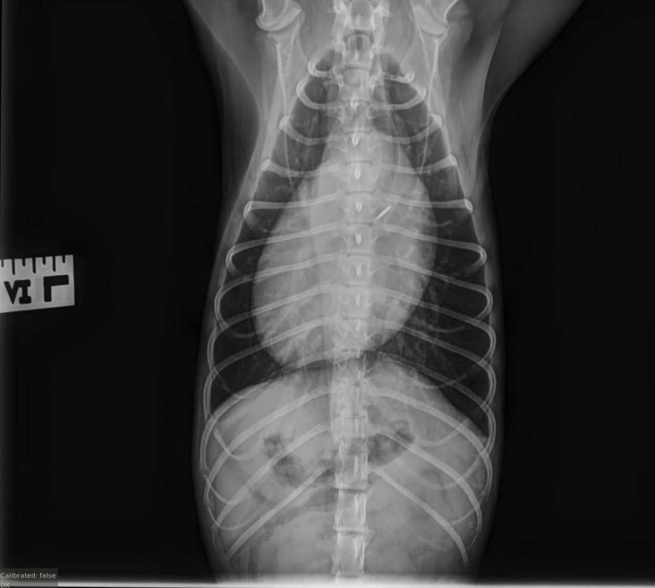
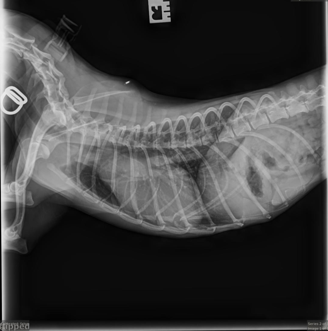
Reference radiographs show the trachea displaced dorsally, consistent with left ventricular enlargement. Also, there is loss of the caudal cardiac waist, consistent with left atrial enlargement. The aorta is severely dilated. The pulmonary parenchyma is normal. The pulmonary vessels are normal. The pleural space is normal.
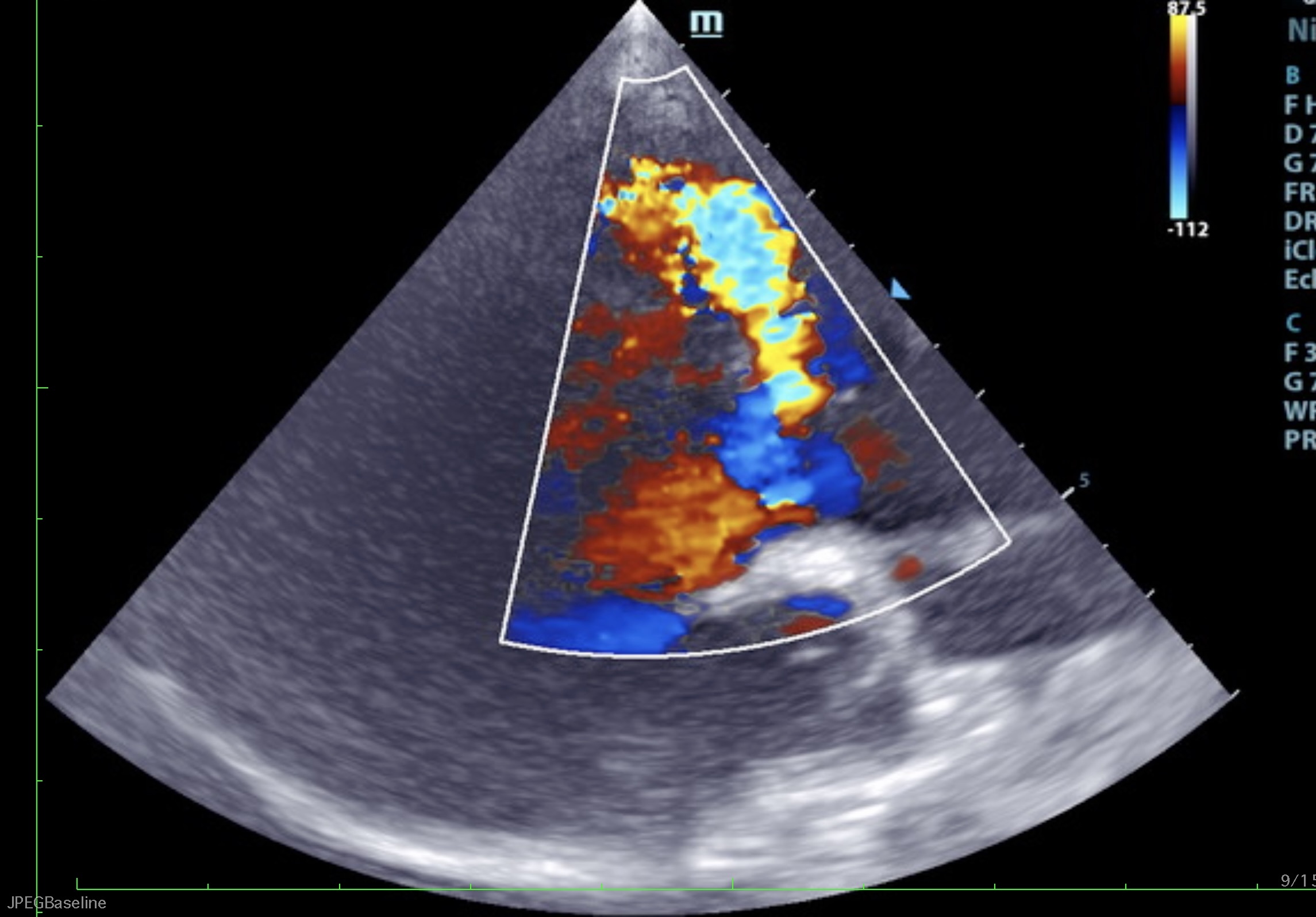
Short axis view of the ventricular septal defect with left to right flow, SDEP® position 1
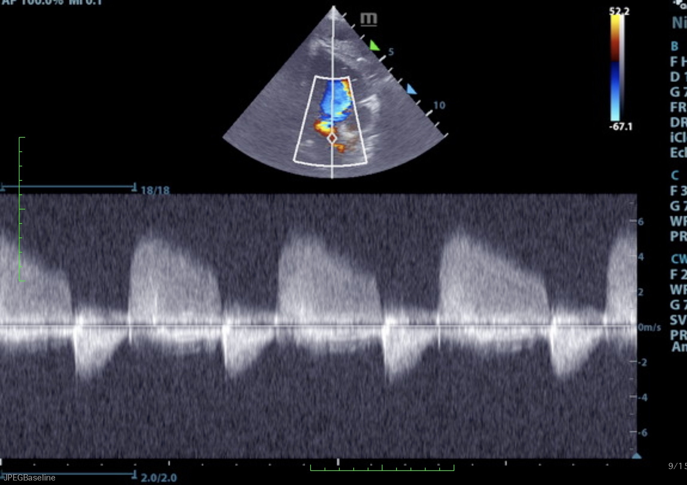
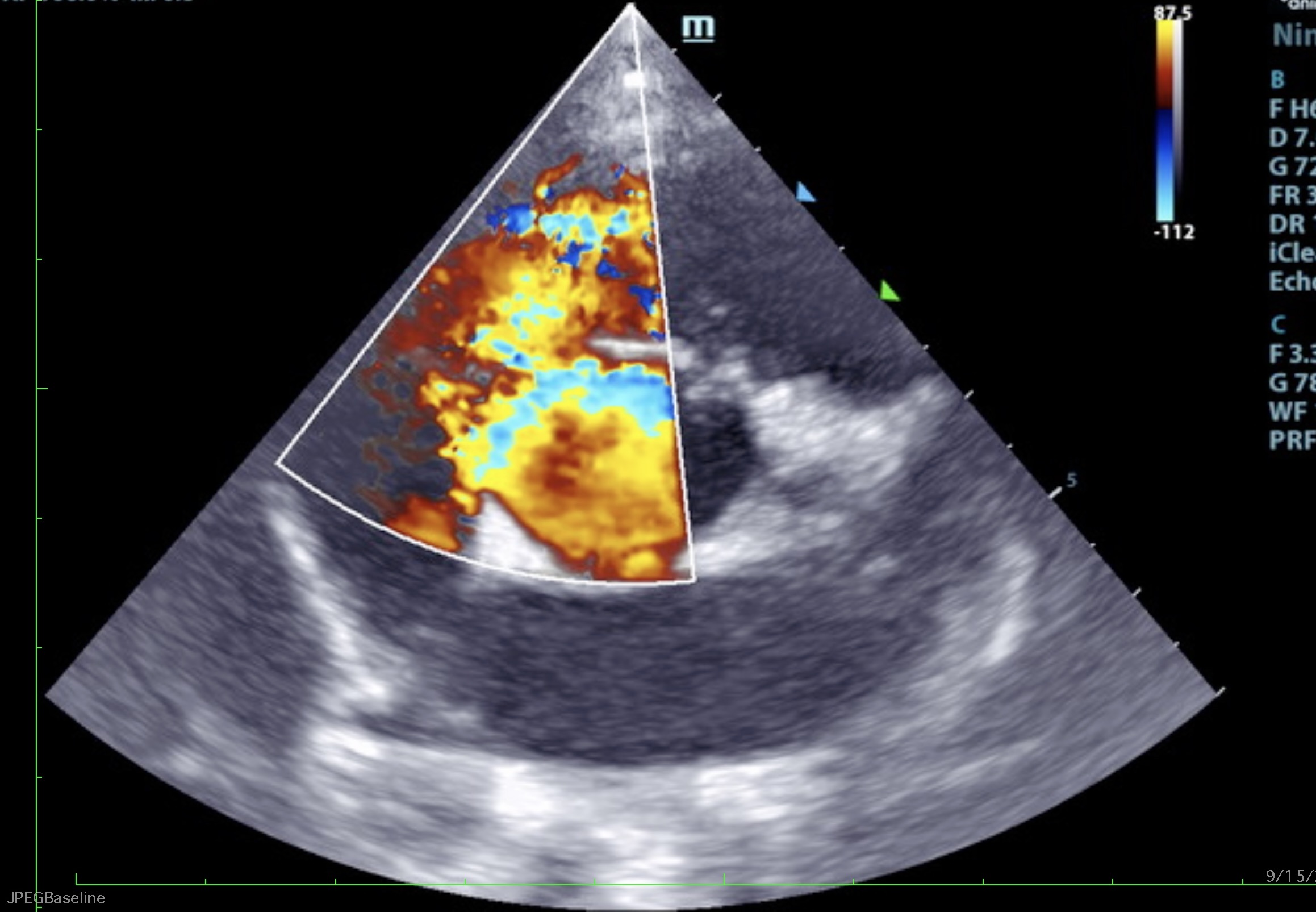
Long axis view of the ventricular septal defect with left to right flow, SDEP® position 1
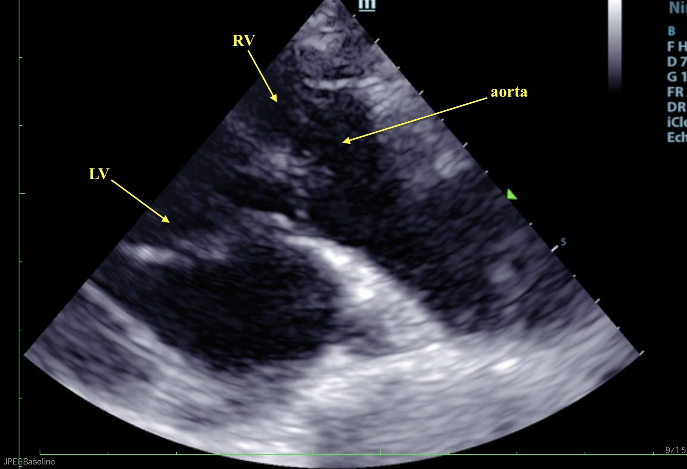
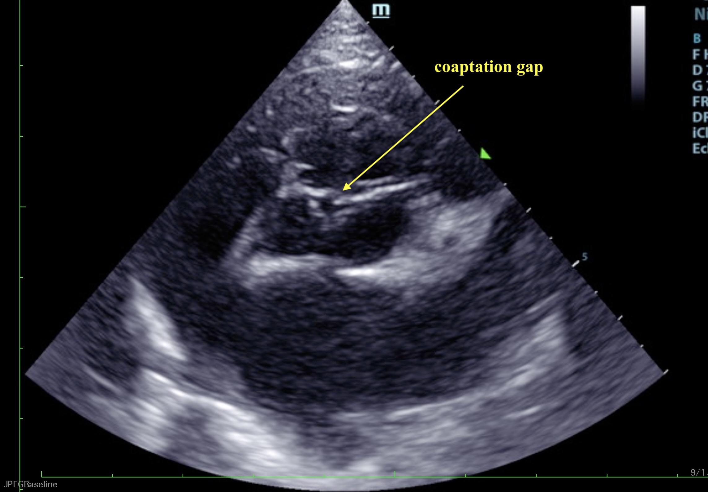
Short axis view of the dilated aorta and aortic valve with central coaptation gap, SDEP® position 1
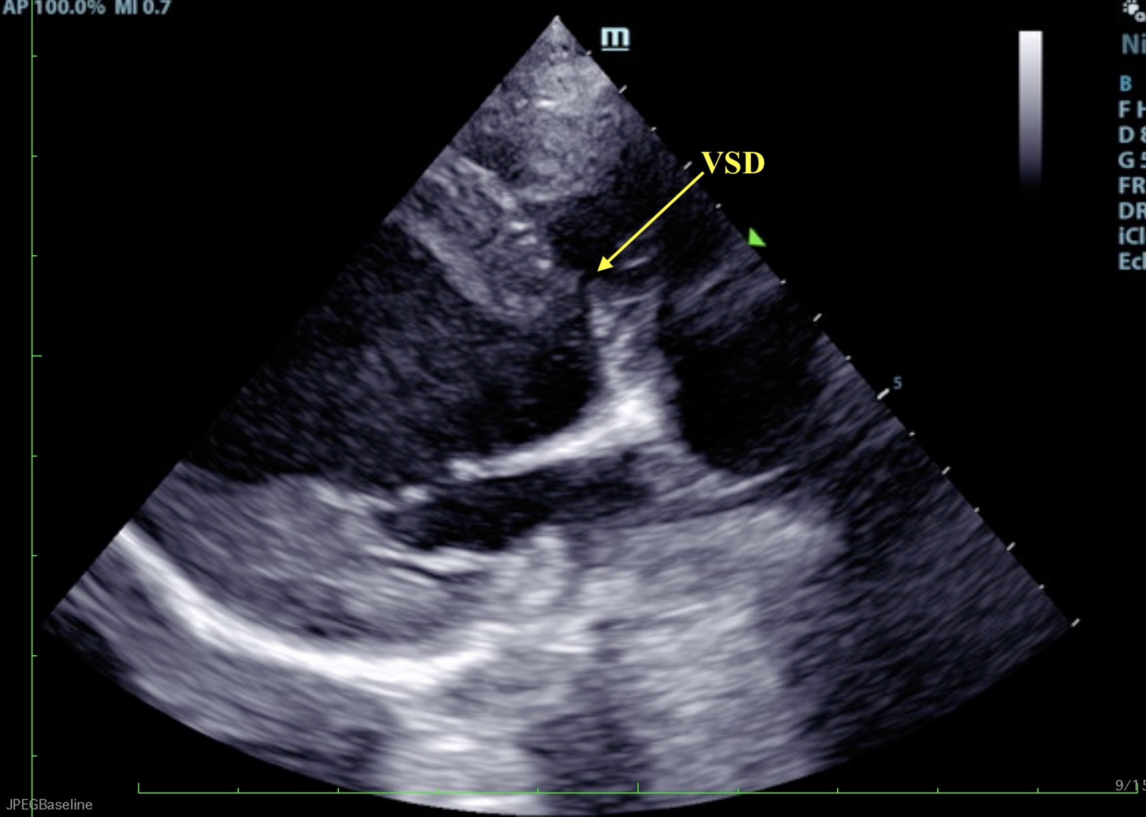
Long axis view of the ventricular septal defect and overriding aorta, SDEP® position 1
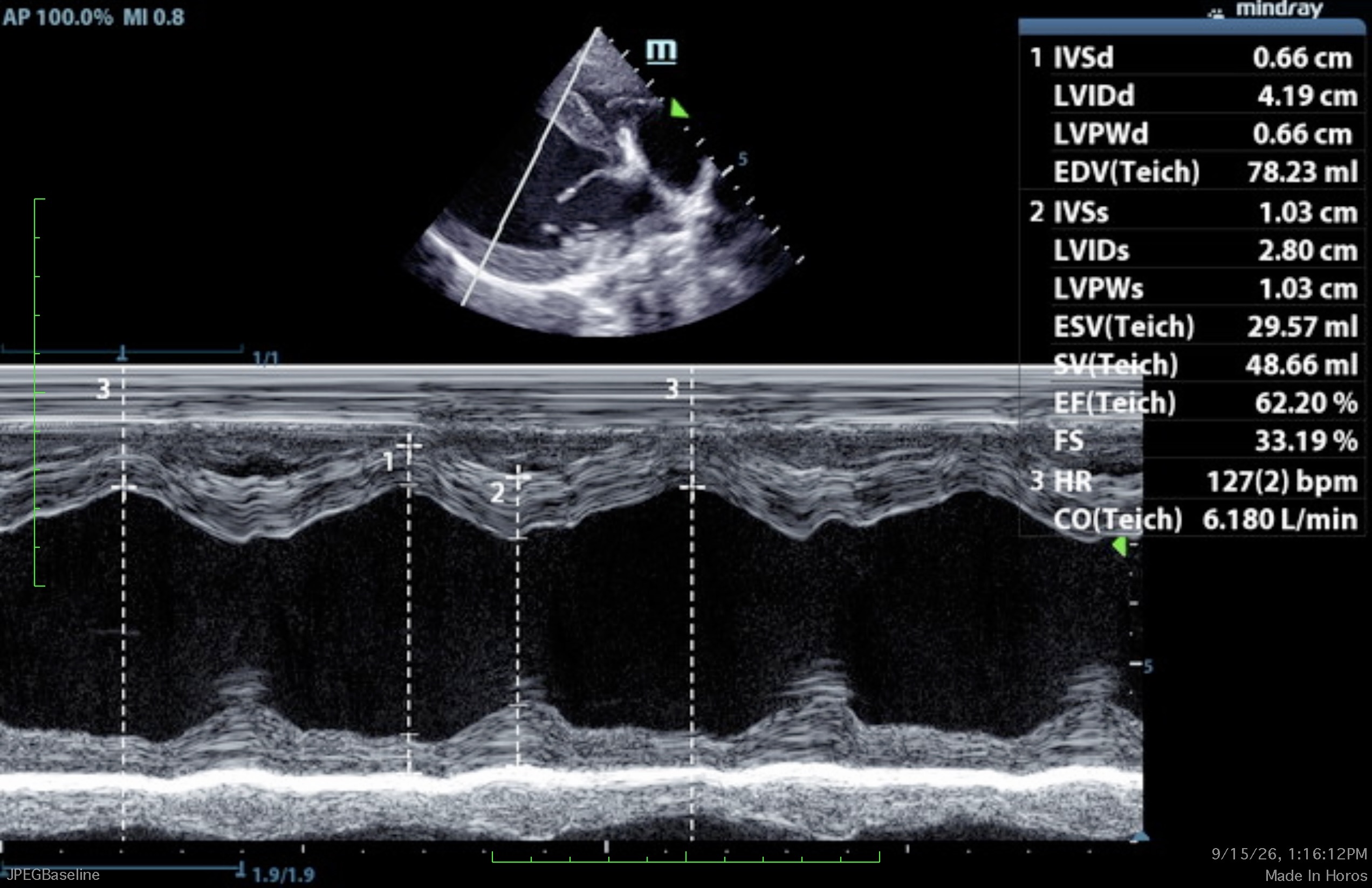
The left atrium is normal in dimension. The left ventricle is severely enlarged, with severely reduced systolic function. The right atrium and ventricle are normal in dimension, with normal systolic function.
The anterior and posterior mitral valve leaflets are appropriately thin with adequate apposition, intact chordae, and there is no significant prolapse. There is no significant mitral regurgitation identified. The tricuspid valve leaflets are appropriately thin with adequate apposition, intact chordae, no significant tricuspid regurgitation, and no evidence of pulmonary hypertension. The left ventricular outflow tract demonstrated severely turbulent flow with an overriding aorta and coaptation gap of the aortic valve leaflets. The left ventricular outflow tract velocity is marginally increased. There is a ventricular septal defect with high velocity left-to-right systolic flow documented. The right ventricular outflow tract assessment revealed laminar flow, with appropriate main pulmonary artery diameter and right pulmonary artery distensibility. The right ventricular outflow tract velocity is normal. There is mild pulmonic and severe aortic valve insufficiency identified. There is no visible pericardial, pleural, or free peritoneal fluid documented.
No evidence of hepatic venous congestion is noted. The cardiac chambers, pericardial and visible extra-cardiac regions were free of masses, spontaneous echo contrast, and thrombi.
- Overriding aorta with a resulting ventricular septal defect (VSD)
- Suspected (no ECG attached) high left-to-right systolic flow across the VSD, with severe aortic regurgitation as a result in the coaptation gap between aortic valve leaflets
- Severely enlarged left ventricle with reduced systolic function
Given the degree of left ventricular dilation, Vetmedin (pimobendan) (0.25–0.35 mg/kg BID) and enalapril (0.5mg/kg) BID) are recommended. Baseline chemistry panel and thoracic radiographs are recommended now, and again with a blood pressure in 1–2 weeks. Referral to a veterinary cardiologist is highly encouraged for reevaluation of the defect.
This patient was scheduled with a veterinary cardiologist immediately. The cardiologist agreed with the findings of overriding aorta and severe VSD. Enalapril was to be continued at the same dose; pimobendan was increased to 1.25 mg q 12 hours. Furosemide was indicated for his recent cough, and spironolactone was started.
Plan:
- Follow the provided list of signs to monitor
- Continue medications
- Recheck radiographs in 3–6 months
- Hematocrit every 3 months to monitor for left-to-right shunting
- Recheck in 4 months and discuss neutering at that point
This patient has been adopted!
Discover SonoPath’s Educational Telemedicine® and Internal Medicine Teleconsultation Services:
Starting on page 413, you can read a whole chapter on PDA including its clinical signs, clinical findings & diagnostics, and treatment suggestions.
Check out the section: Ventricular Septal Defect (pg 456-462) for a closer look at a case like this.

Approximate Study Time: 45 mins – 1 hour (self-paced)
Instructor: Eric Lindquist, DMV (Italy), DABVP (Canine & Feline), Cert. IVUSS, Founder & CEO of SonoPath
Expand your diagnostic awareness with this curated collection of echocardiographic pathology encountered in real-world clinical practice.
The Scope of SDEP® Echo Pathology library presents 247 images illustrating a wide range of cardiac abnormalities seen on a routine basis in veterinary sonography. While no single resource can encompass the full spectrum of cardiac disease, this library is designed to reinforce one of the most important principles in ultrasound: expect the unexpected.
By exposing you to diverse pathological presentations, this collection strengthens your ability to recognize variation, identify abnormalities, and approach each case with a more critical and informed eye. Ideal as both a learning tool and a quick-reference resource, this library supports ongoing development of pattern recognition and diagnostic confidence in echocardiography.

2027 JULY
SonoPath International Summit Halifax
21 RACE-approved CE
Follow the heart to Halifax for three days of focused veterinary cardiology and ultrasound education featuring leading practitioners and educators in the field.
Designed for experienced sonographers, veterinarians, cardiologists, and veterinary professionals with an interest in cardiac imaging, this Summit combines clinically relevant lectures, case-based learning, and expert perspectives in an intimate waterfront setting.
2027 SDEP® Echo Hands-On Ultrasound Training
22 RACE-approved CE credits
Live lectures, hands-on training, & small-group instruction
Designed for veterinarians and veterinary technicians looking to build or advance practical echocardiography skills using a structured, repeatable protocol. Whether you are new to cardiac ultrasound or refining existing skills, the SDEP® Echo Protocol allows personalized training tailored to your experience level. Beginners to echocardiography will build confidence while experienced sonographers refine and develop speed, consistency, and diagnostic accuracy.
2027 FEBRUARY
Specialist Intensive SDEP® Abdominal Sonography & Pathology
14 RACE-approved CE
Live Lectures & 2 scanning sessions
Limited Space Available
Join SonoPath in beautiful Fort Collins, CO at Colorado State University for a small, high-level SDEP® experience built exclusively for boarded and boarding internists. This is where standard scanning stops and intentional, repeatable diagnostic efficiency begins.