Many thanks to Elizabeth Fowler, DVM, CCRP, practice owner and veterinarian at Infinite PawsAbilities located in New Braunfels, TX, for providing the diagnostic ultrasound images for this case and to her staff for their compassionate care of this sweet Siberian Husky. Specialist interpretation by Nele Eley, DVM, Dr. med. vet., DipECVDI, European Specialist in Veterinary Diagnostic Imaging, Cert. Radiology,
Senior Lecturer University of Giessen, Germany, Veterinary Faculty, Department of Radiology. Read on for a stellar example of the diagnostic efficiency of orthopedic ultrasound, when the “normal” cruciate ligament injury was on the menu!

History
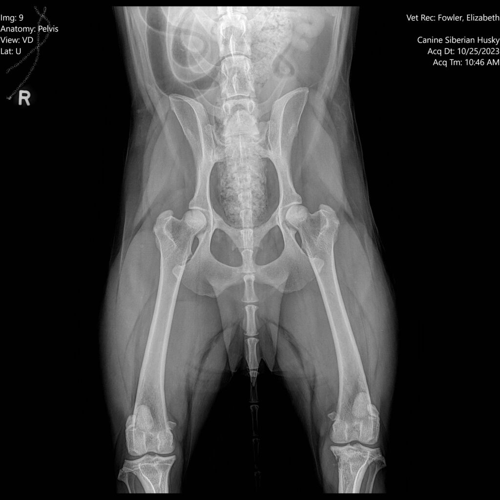
The patient was presented for recent decrease of appetite, reduced activity, and left hind lameness after playing. Physical examination found crepitus and laxity in the left stifle, with some crepitus noted in the right stifle. Weakness in the hind limbs was also noted, with diminished extension in the hips and stifles. Chem 6 was WNL.
Ultrasound Findings
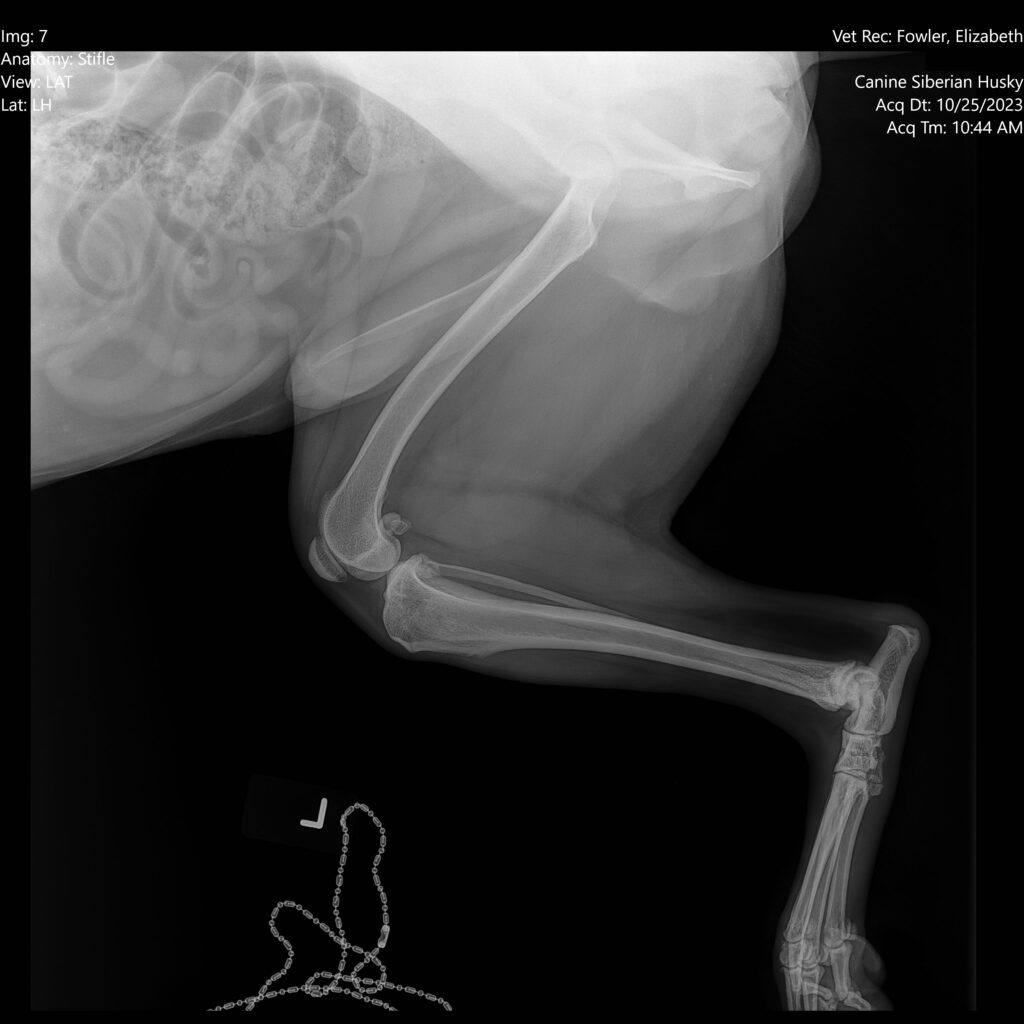
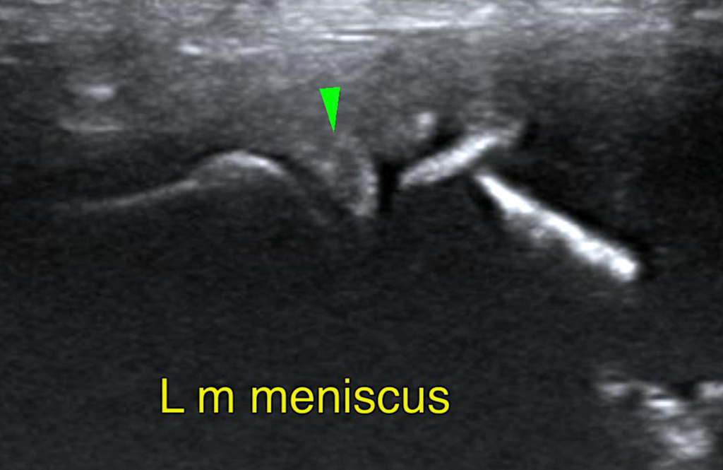
Left Stifle – Scant effusion and minimal periarticular bone remodeling was noted within the left stifle joint. There was no evidence of synovial or capsular thickening or proliferation. The cranial cruciate ligament appeared to be continuous and well-delineated, no deviation from normal echo architecture was noted. Lateral and medial menisci were within their anticipated positions and aligned well below the bone surfaces; meniscal surfaces were even and smooth. The echotexture was hypoechoic and uniform. The joint margins were smooth, no osteophytes were seen. The infrapatellar fat pad was present with the expected echo architecture.
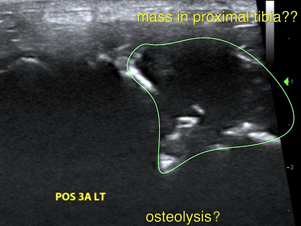
There appeared to be a cortical bone defect in the medial contour of the left tibia with a hypoechoic heterogeneous mass expanding from it.
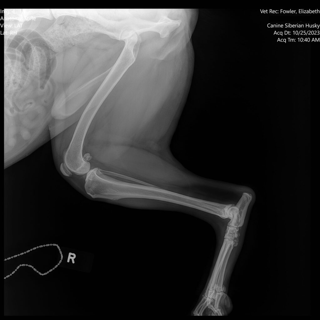
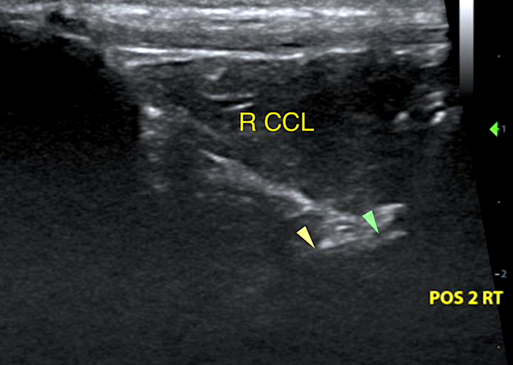
Right Stifle – Scant effusion and minimal periarticular bone remodeling was noted within the right stifle joint. There was no evidence of synovial or capsular thickening or proliferation. The cranial cruciate ligament appeared to be continuous and well-delineated, no deviation from normal echo architecture was noted. Lateral and medial menisci were within their anticipated positions and aligned well below the bone surfaces; meniscal surfaces were even and smooth. The echotexture was hypoechoic and uniform. The joint margins were smooth, no osteophytes were seen. The infrapatellar fat pad was present with the expected echo architecture.
Diagnosis
Normal age-related presentation of the stifle joint with no evidence of cruciate ligament or meniscal injury. Aggressive osteolytic lesion in the left proximal tibia is suspected.
Interpretation of the Findings
The stifle joints presented within age-related normal limits. There is no evidence of cruciate ligament or meniscal injury noted in the ultrasonographic study. The ultrasonographic presentation of the left proximal tibia, however, is abnormal. Potential for an aggressive osteolytic lesion in the left proximal tibia is suspected based on the images.

Further Recommendations
Ultrasonographic recheck with eventual sampling of the soft tissue component of the suspected mass could be considered alternatively. The patient’s treatment plan included a consultation with an orthopedist/oncologist where a CT scan will be performed. Depending on those findings, most likely amputation of the left hind leg will be the outcome.
Current Case Status
The patient went on to receive a CT scan which showed that there was a fracture of the left tibia, as well as metastatic cancer in his lungs. Amputation of the left leg was performed, and the patient is receiving radiation and rehabilitation therapy at Texas A&M. The patient was still experiencing loss of appetite and had some liver elevations. After being treated with Entyce for a few days, the patient’s appetite returned and he is now on Denamarin. Patient has been doing okay since.