
Yes, cats get heart base masses too
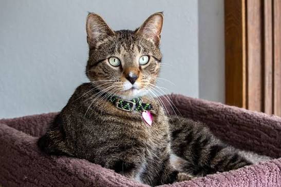
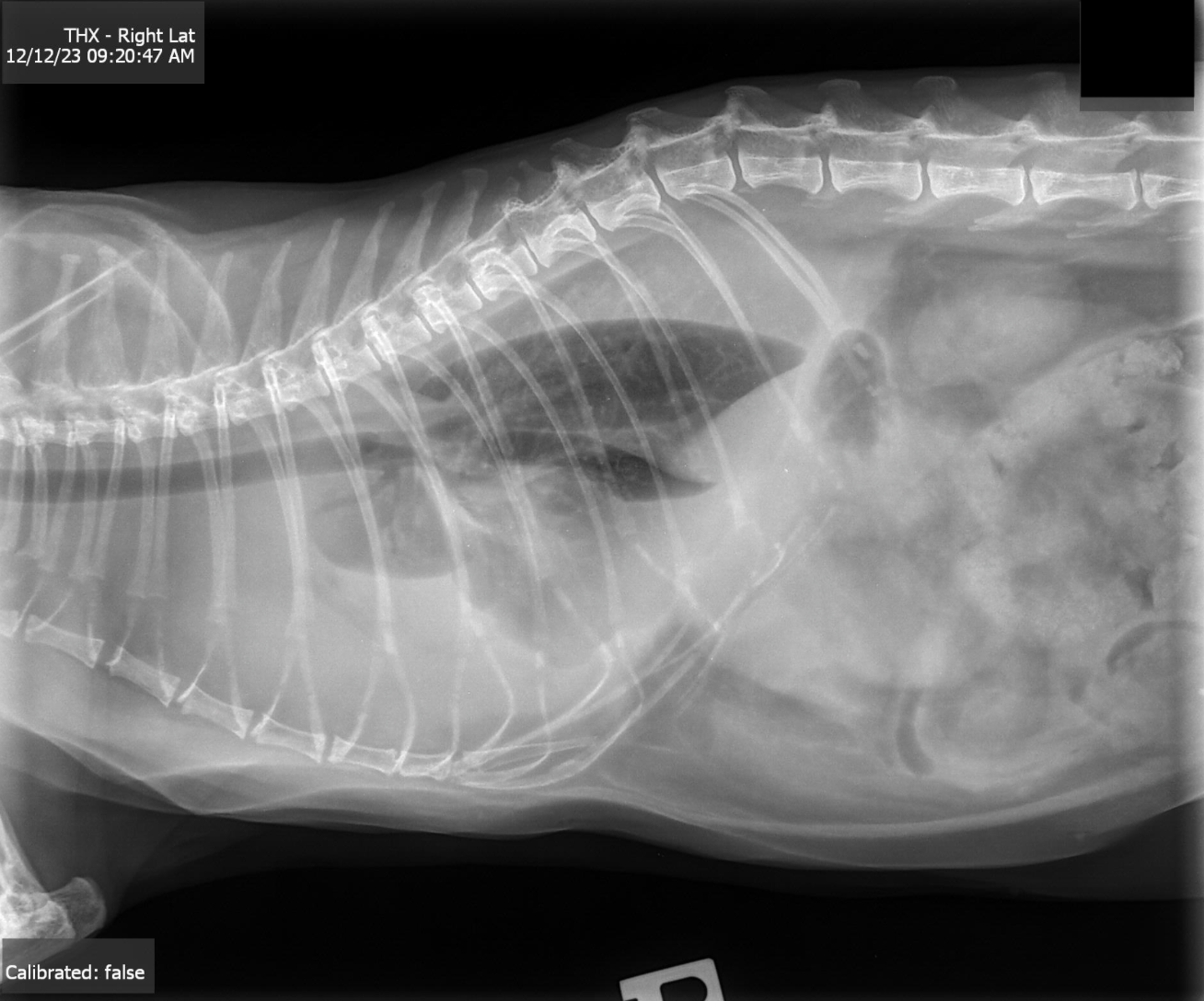
A feline patient presented to Dr. Veysey at Main Street Animal Hospital for a 3-day history of increased respiratory effort, open mouthed breathing, lethargy and decreased appetite. On exam, bronchovesicular sounds were only audible in the dorsal thorax. No heart murmur was appreciated. Thoracic radiograph showed significant pleural effusion obscuring the heart. The patient was started on Lasix and an echocardiogram was recommended.
Sonographer: Crystal Hill, RVT, The Focal Zone
Specialist: R. McKenzie Daniel, DVM, DABVP (Canine and Feline)
Referring Veterinarian: Paige Veysey, DVM, Main Street Animal Hospital, Ontario
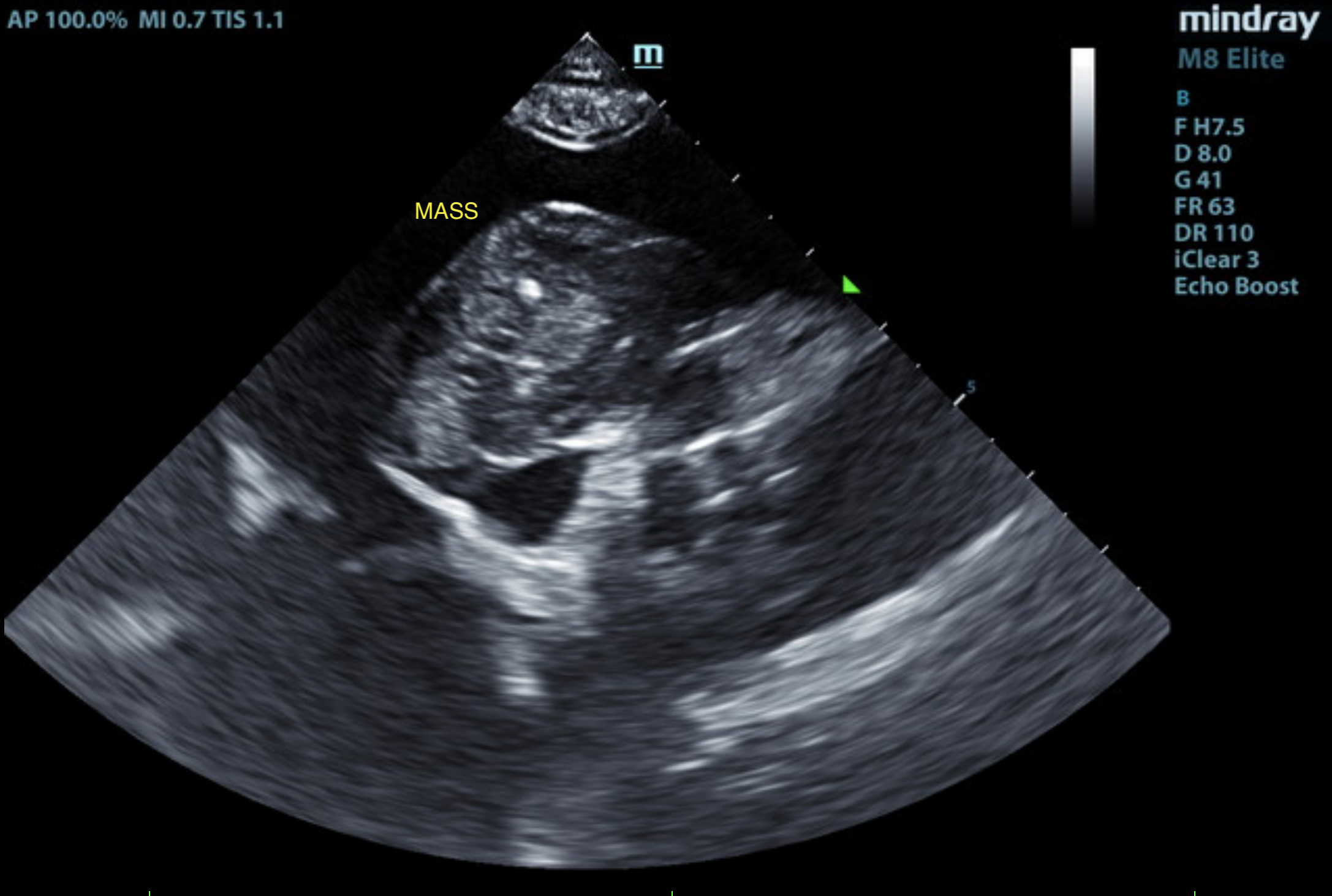
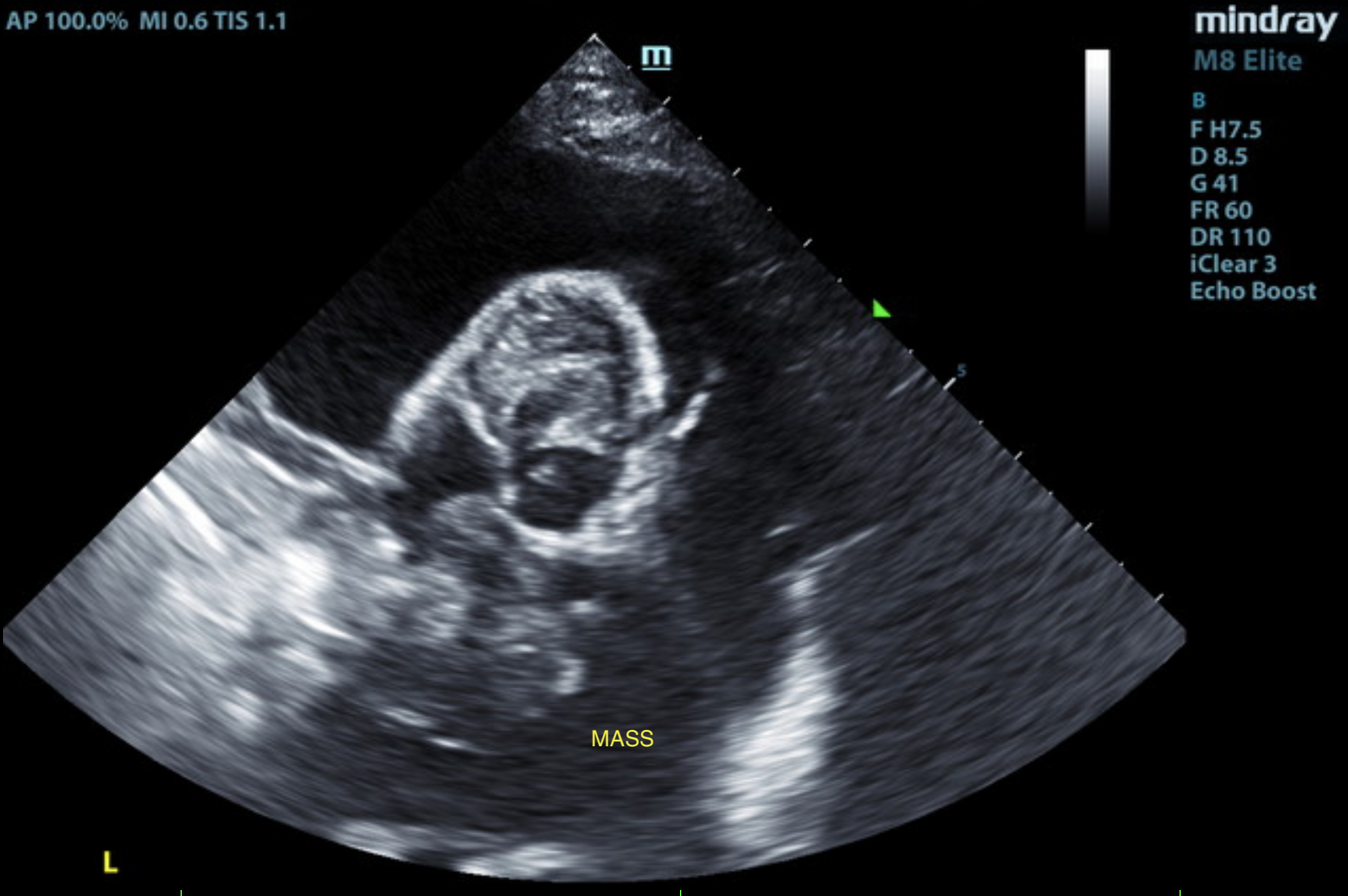
US Video at SDEP® Position 1 going in between the 4 and 5 chamber views with mass invading into right atrium.
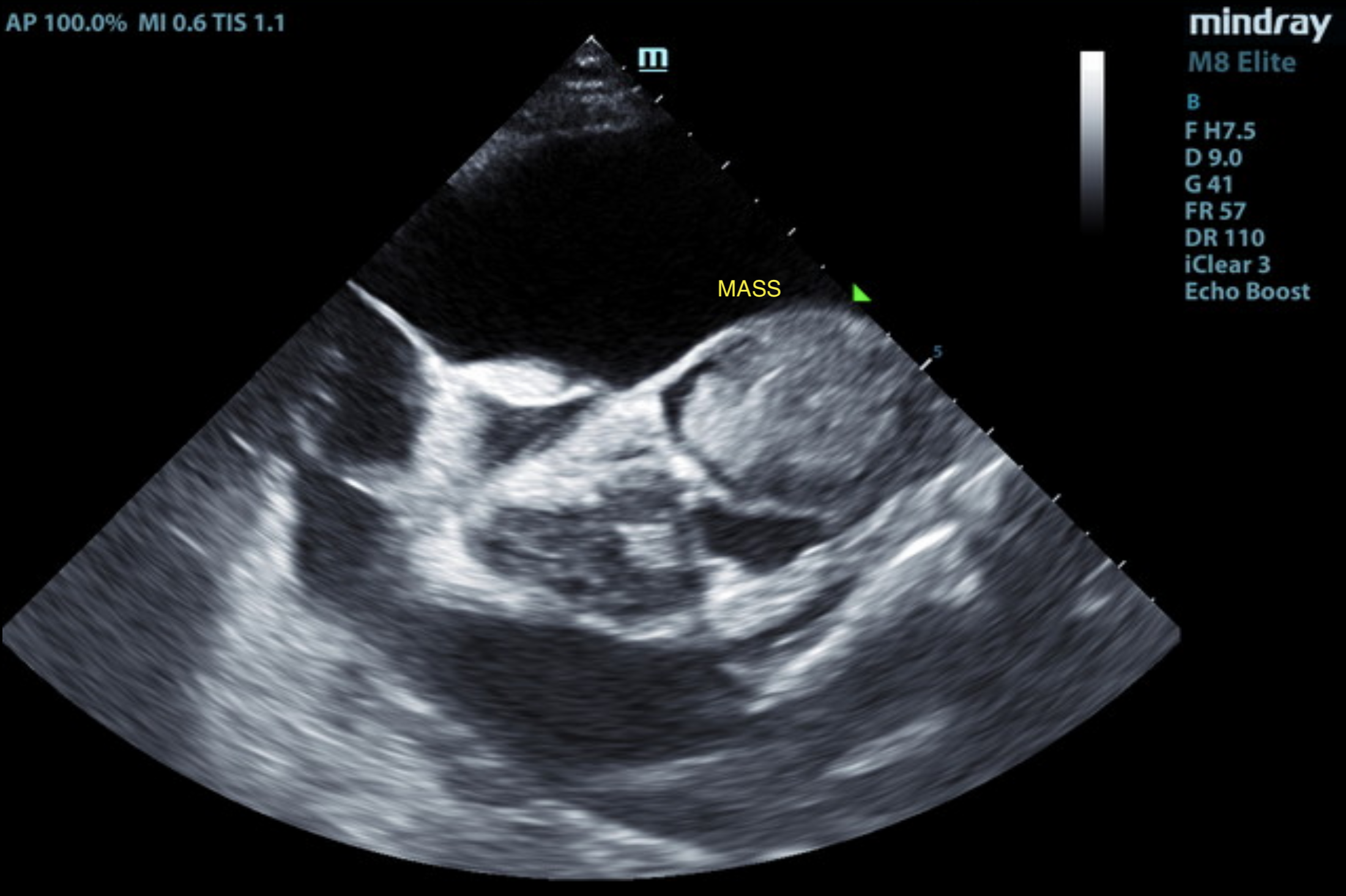
US video SDEP® Position 2 showing the short axis view at the level of the aorta and left atrium with mass present at heart base.
US video SDEP® Position 1 showing the 4 chamber view, mass, and color doppler.
The echocardiogram in this patient demonstrated normal left atrial size based on two separate LA measurements. The cranial and caudal mitral valve leaflets presented normal linear structure and kinetics. The left ventricle presented normal thicknesses with primarily maintained linear contour and reduced LV volume. The myocardium presented normal echogenicity without subjective evidence of significant fibrotic or ischemic disease. The contractility of the ventricular walls was adequate and in normal range for this patient evidenced by the fractional shortening measurement and subjective evaluation of the different regions and angles of the myocardium. The left ventricular outflow tract demonstrated normal laminar flow and subjective structural integrity. The right atrium and auricle revealed normal size, structure and content. Tricuspid valvular assessment demonstrated adequate linear morphology and kinetics. The right ventricle was of normal size (1/3 diameter of LV), chordae structure, myocardial echogenicity and thickness. Pulmonic tract assessment revealed normal valve structure, laminar flow, and diameter (approx.1:1 PA/AO ratio). No overt pericardial free fluid was noted with moderate volume; primarily anechoic free pleural fluid present.
A non-homogeneous focally hyperechoic mass was present in the area of the heart base measuring 1.3-3.0 cm in diameter.
• Non-homogeneous focally hyperechoic cardiac vs pericardial pulmonary mass area of heart base.
• Moderate non-cardiogenic pleural effusion.
Unfortunately, the cardiac vs pericardial pulmonary mass is most consistent with neoplastic criteria exhibiting potential for focal mass mineralization. Effusion analysis cytology +/- C/S if evidence of inflammatory cells could be considered in search for definitive diagnosis. However, an unfavorable prognosis is indicated.
Cardiac neoplasia is very rare in cats. The most common types of cardiac neoplasias reported are hemangiosarcomas, chemodectomas, lymphomas, and ectopic thyroid carcinomas. In cats these neoplasms are usually primary, malignant and the majority are extranodal lymphomas. Most secondary/metastatic tumors are found in the left ventricular free wall and interventricular septum.
Patients with a cardiac mass can present with no clinical signs or they can present with signs of respiratory distress and decreased cardiac output, as well as signs of right sided congestive heart failure related to pleural and pericardial effusions from the mass. Clinical signs include acute collapse, exercise intolerance, lethargy, weight loss, vomiting, syncopal episodes or sudden death. On physical examination there may be muffled heart sounds, tachycardia, pulse deficits, pale mucous membranes, weak femoral pulses, ascites, and tachypnea/dyspnea.
Cardiac neoplasms may be detected on radiographs but typically ultrasound is needed to confirm. An echocardiogram can determine location and size of mass and give some insight into the type of mass. Definitive diagnosis is challenging as cardiac masses are not routinely sampled due to the risk of arrhythmias and hemorrhage. More commonly a pleural or pericardial effusion is sampled to determine etiology. Necropsy and histopathology are utilized for definitive diagnosis post mortem.
Treatment in these cases is challenging as it is difficult to obtain a definitive diagnosis. Surgery, chemotherapy, radiation therapy, and supportive treatment such as pericardio/thoracocentesis can help extend life but in general the outcome is usually poor. In cats diagnosed with lymphoma, some are able to achieve remission using the CHOP protocol. 1 2 3
The patient continued to decline and was humanely euthanized.
14 RACE-approved CE credits
On-demand webinar lecture series plus post-course CE quizzes
Approximate Study Time: 14+ hours (self-paced)
Instructor: Peter Modler, DVM, Dipl.-Tzt, Specialist German Board
Build confidence and clarity in cardiology with this comprehensive, clinically focused lecture series led by Dr. Peter Modler, a leading expert in mitral valve repair. Designed to make cardiology logical and approachable, this program walks you through essential principles from basic hemodynamics and physiology to advanced echocardiographic interpretation and case-based decision-making.
6 RACE-approved CE credits
On-demand lectures and step-by-step technique videos plus post-course CE quizzes
Approximate Study Time: 6–8 hours
Instructor: Eric Lindquist, DMV (Italy), DABVP (Canine & Feline), Cert. IVUSS, Founder & CEO of SonoPath
Build confidence in echocardiography with a structured, step-by-step protocol designed to make cardiac ultrasound approachable, repeatable, and clinically relevant. The SDEP® Echo progression uses a proven numbering system, just 7 maneuvers across 4 key points, to guide you through a complete and efficient cardiac exam.

November 13-14, 2026
16 RACE-approved CE
Designed for advanced veterinary sonographers ready to refine echocardiographic technique, improve diagnostic interpretation, and integrate advanced cardiac pathology into clinical decision-making.

2027 SDEP® Echo Hands-On Ultrasound Training
22 RACE-approved CE credits
Live lectures, hands-on training, & small-group instruction
Designed for veterinarians and veterinary technicians looking to build or advance practical echocardiography skills using a structured, repeatable protocol. Whether you are new to cardiac ultrasound or refining existing skills, the SDEP® Echo Protocol allows personalized training tailored to your experience level. Beginners to echocardiography will build confidence while experienced sonographers refine and develop speed, consistency, and diagnostic accuracy.
SDEP® Echo REMOTE Live Virtual Training Weekend
22 RACE-approved CE credits
Join us remotely with a direct Zoom link to your SDEP® instructor, you scan from your home machine on your patients training with the watchful support of not only your instructor, but a dedicated breakout room instructor.
Check out the Curbside Guide chapter 8 “Pericardial Effusion and Cardiac Neoplasia” (pg 369-383) and scan the QR Codes to watch video case studies.

References
1. Aupperle H, März I, Ellenberger C, et al. Primary and secondary heart tumours in dogs and cats. J Comp Pathol 2007;136:18–26.
2. Treggiari E, Pedro B, Dukes-McEwan J, et al. A descriptive review of cardiac tumours in dogs and cats. Veterinary and Comparative Oncology 2017;15:273–288.
3. Amati M, Venco L, Roccabianca P, et al. Pericardial lymphoma in seven cats. J Feline Med Surg 2014;16:507–512.